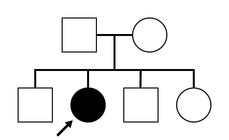
SR1036
AR Cone and Cone Rod Dystrophy (IA1biii)
Female
Female
SR1036
AR Cone and Cone Rod Dystrophy (IA1biii)
Female
Female
Visit at age: 38 years
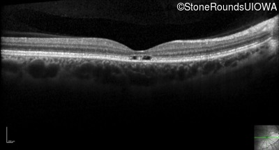
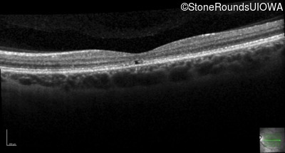
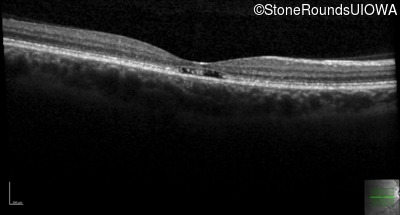
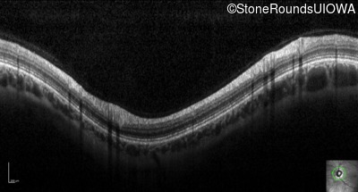
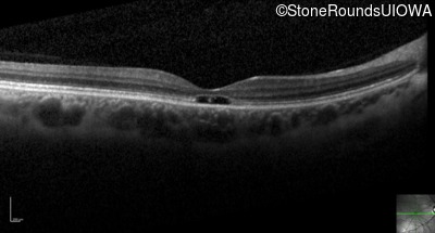
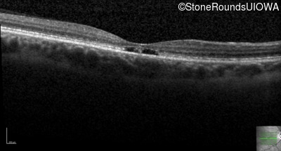
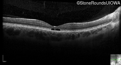
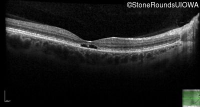
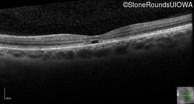
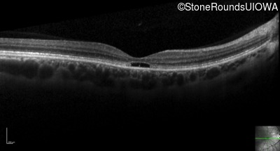
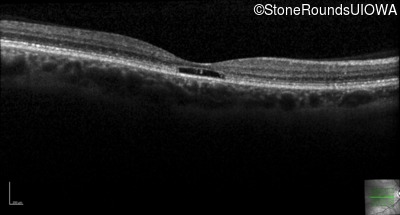
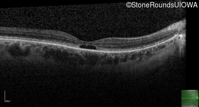
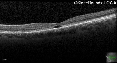
Optical Coherence Tomography - Right - 20/20 -1
Exemplar / OCT Stack
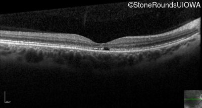
OCT Stack
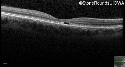
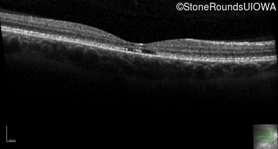
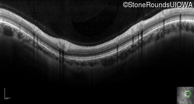
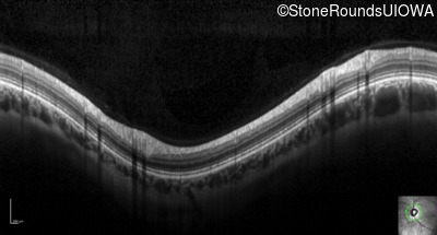
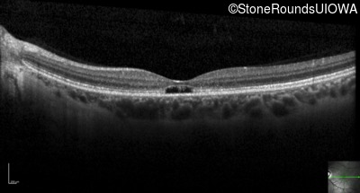
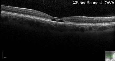
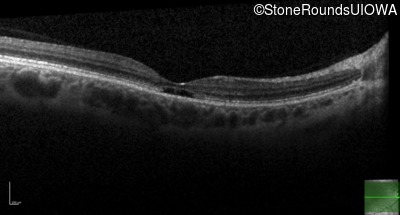
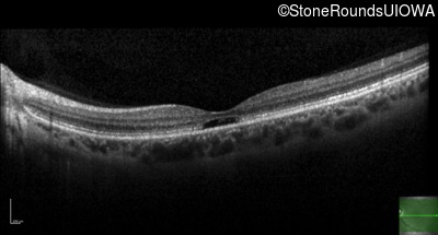
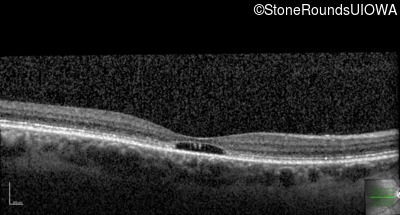
Optical Coherence Tomography - Left - 20/40
Exemplar / OCT Stack
OCT Stack
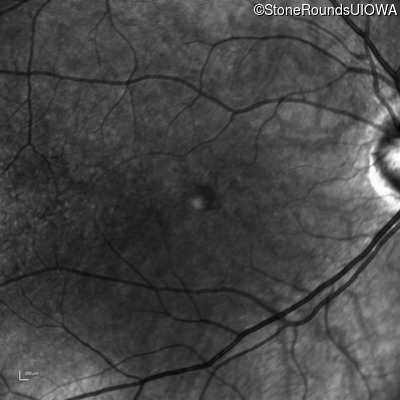
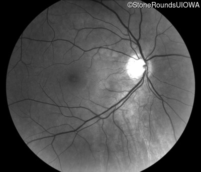
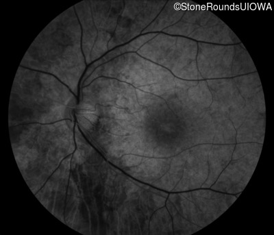
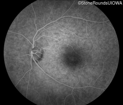
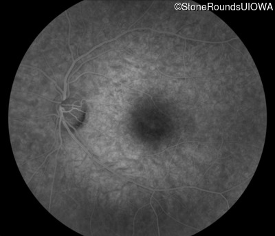
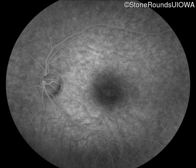
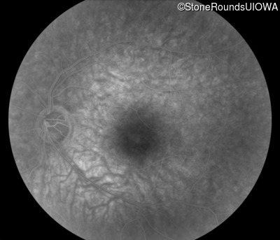
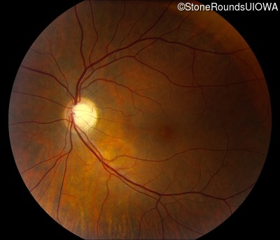
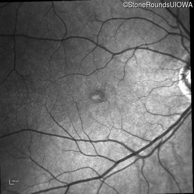
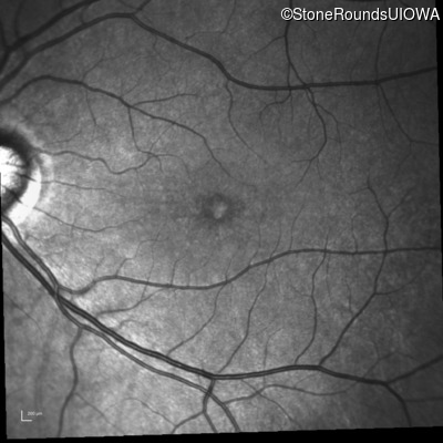
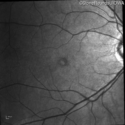
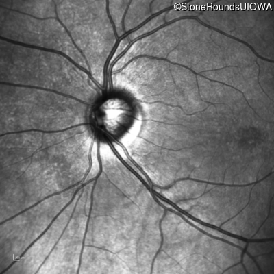
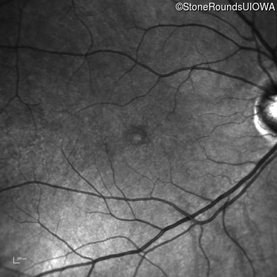
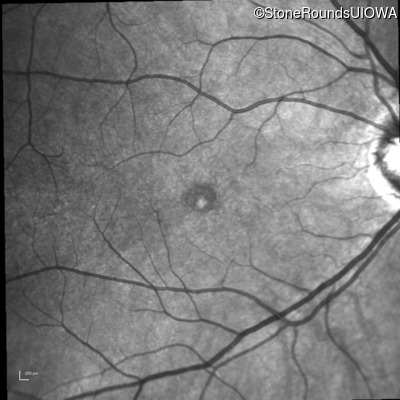
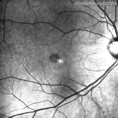
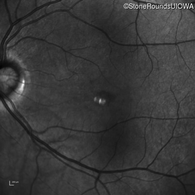
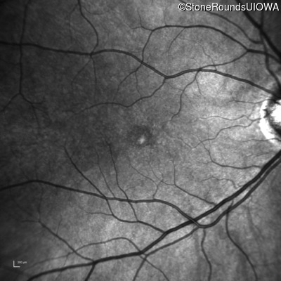
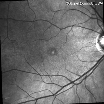
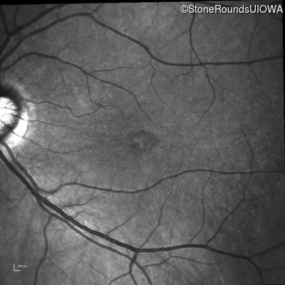
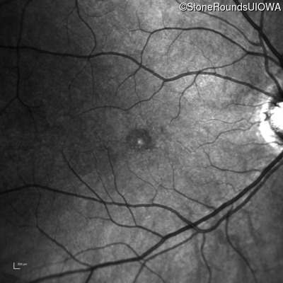
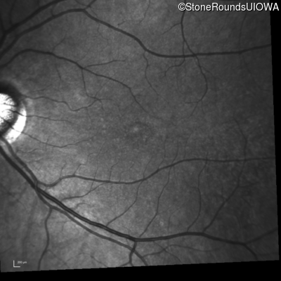
Infrared Fundus Photograph - Right - 20/20 -1
Exemplar
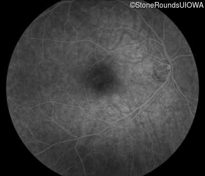
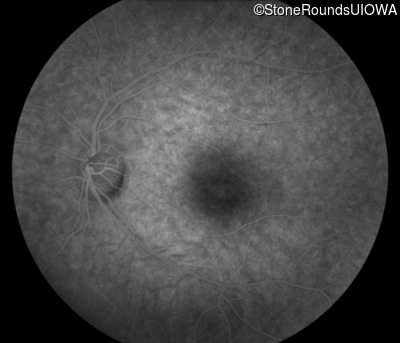
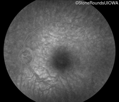
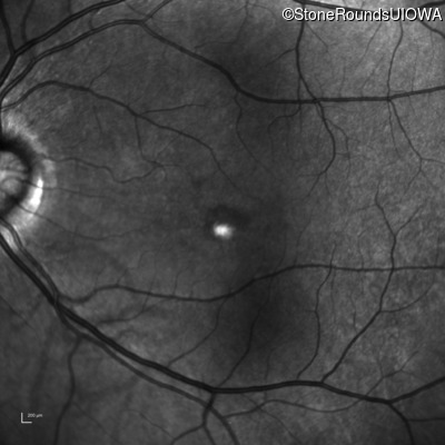
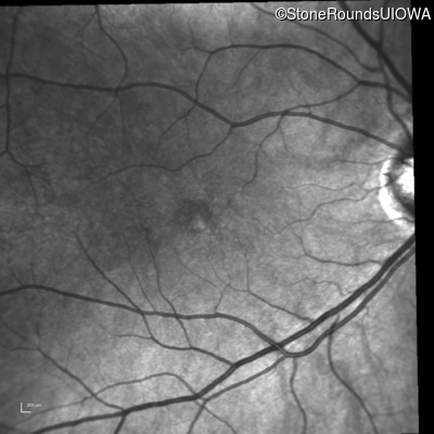
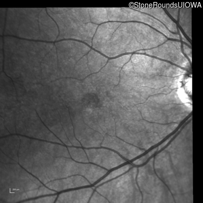
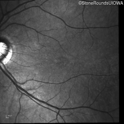
Infrared Fundus Photograph - Left - 20/40
Exemplar
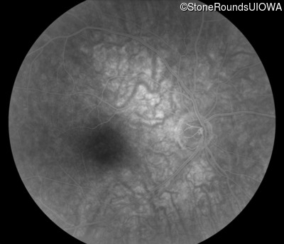
Fluorescein Angiography - Right - 20/20 -1
Exemplar

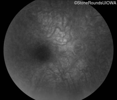
Fluorescein Angiography - Left - 20/40
Exemplar


Visit at age: 38 years (Visit 2)
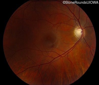
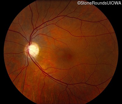
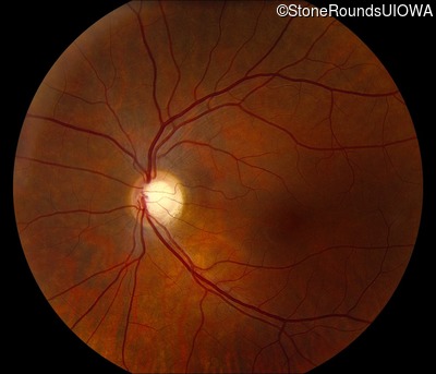
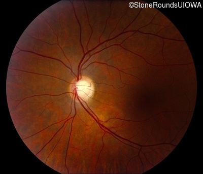
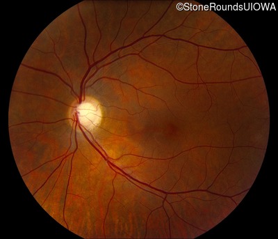
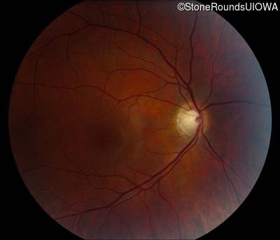
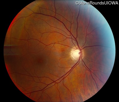
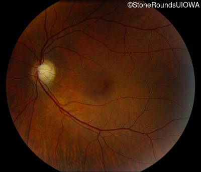
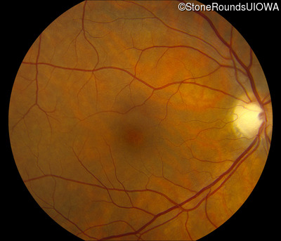
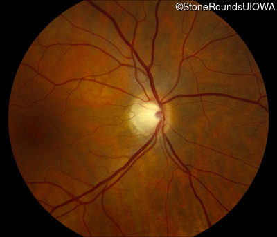
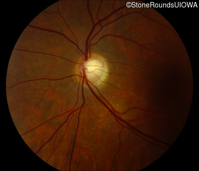
Fundus Photography - Right - 20/25 +1
Exemplar

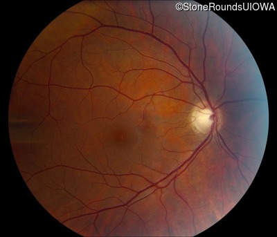
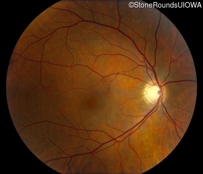
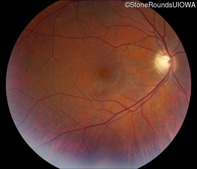
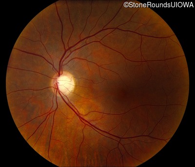
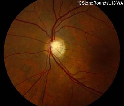
Fundus Photography - Left - 20/32 -1
Exemplar

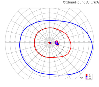
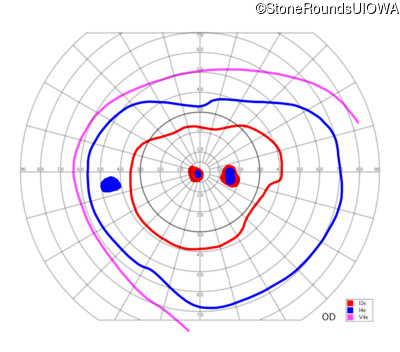
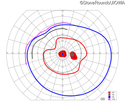
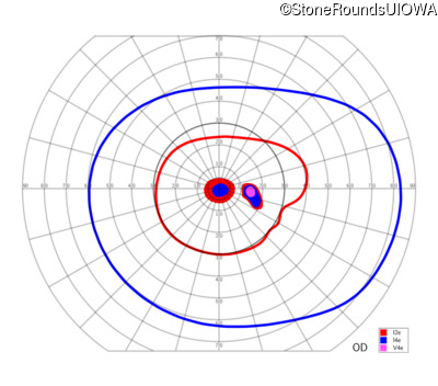
Goldmann Visual Field - Right - 20/25 +1
Exemplar
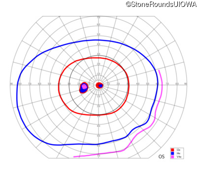
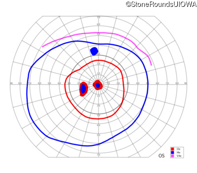
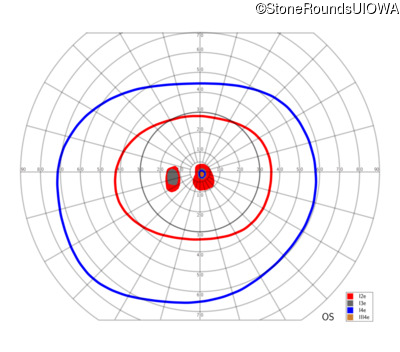
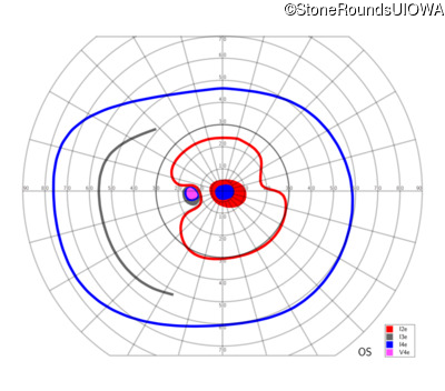
Goldmann Visual Field - Left - 20/32 -1
Exemplar
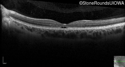
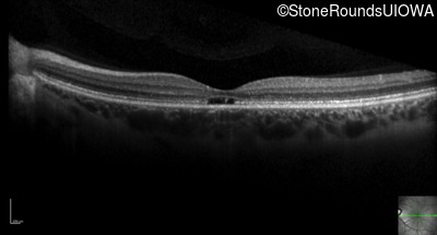
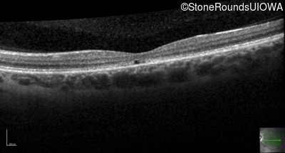
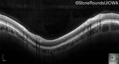
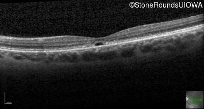
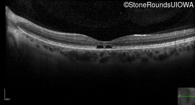
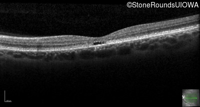
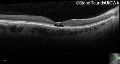
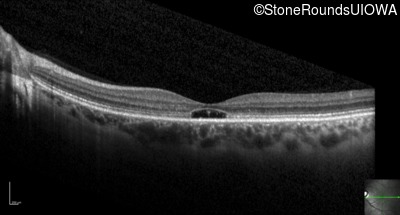
Optical Coherence Tomography - Right - 20/25 +1
Exemplar / OCT Stack
OCT Stack
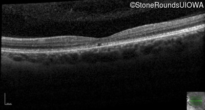
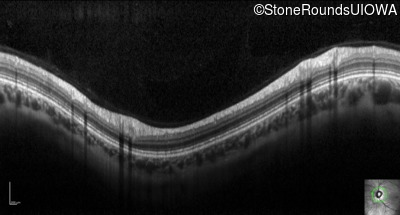
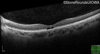
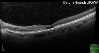
Optical Coherence Tomography - Left - 20/32 -1
Exemplar / OCT Stack
OCT Stack
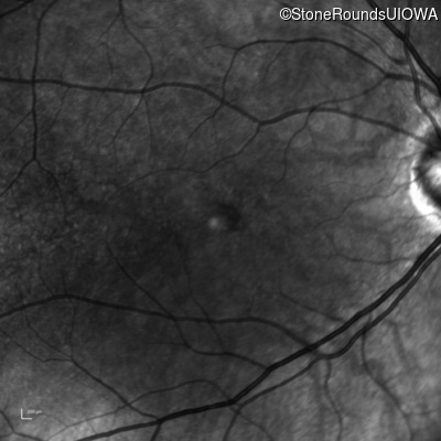
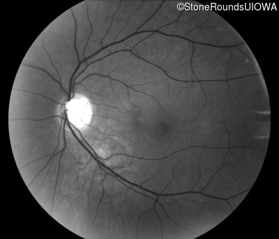
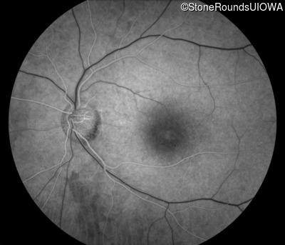
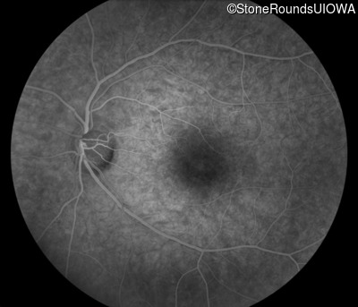
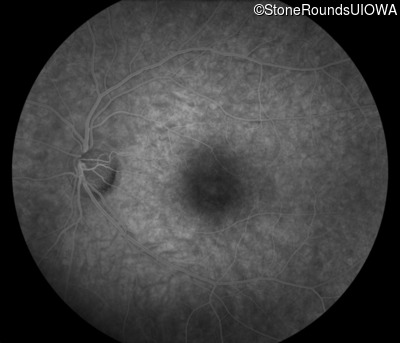
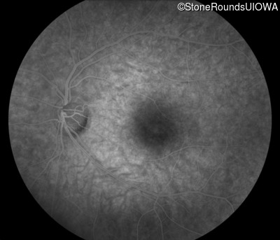
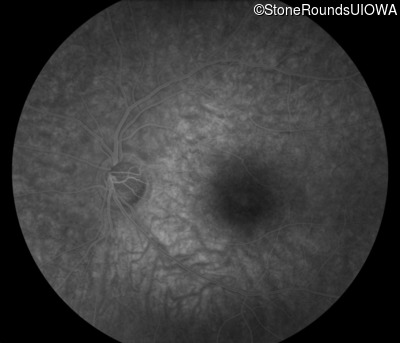
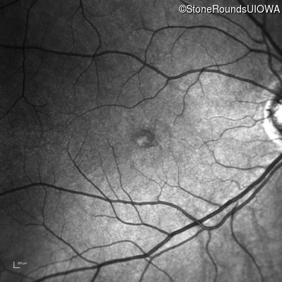
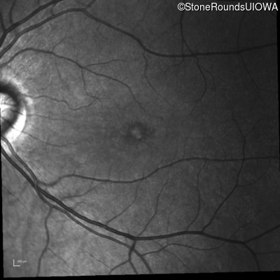
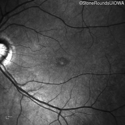
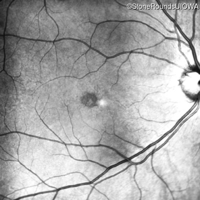
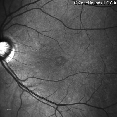
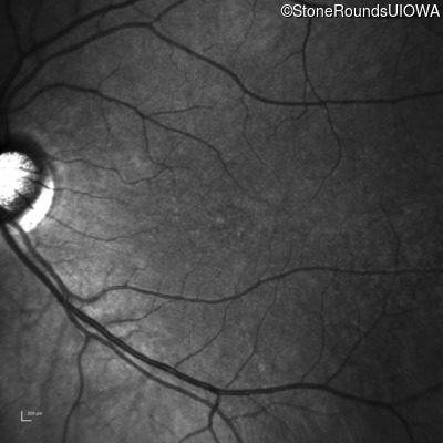
Infrared Fundus Photograph - Right - 20/25 +1
Exemplar
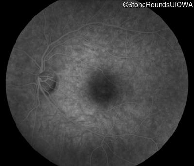
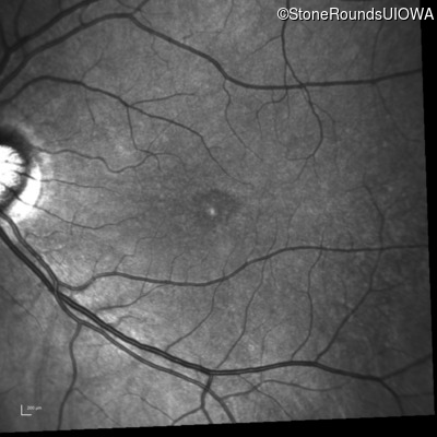
Infrared Fundus Photograph - Left - 20/32 -1
Exemplar
Visit at age: 39 years
Optical Coherence Tomography - Right - 20/25
Exemplar / OCT Stack
Optical Coherence Tomography - Left - 20/50 -2
Exemplar / OCT Stack
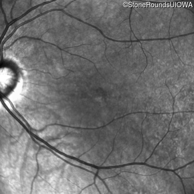
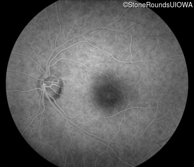
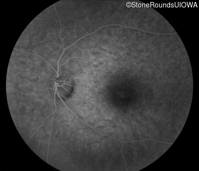
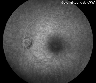
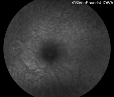
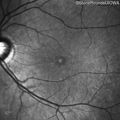
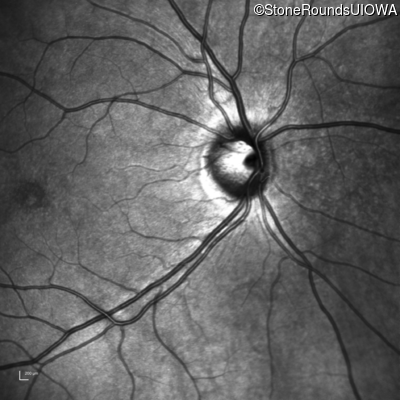
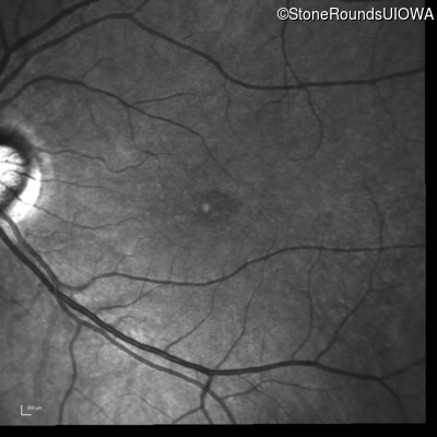
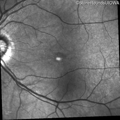
Infrared Fundus Photograph - Right - 20/25
Exemplar
Infrared Fundus Photograph - Left - 20/50 -2
Exemplar


Visit at age: 41 years
Goldmann Visual Field - Right - 20/32 -2
Exemplar
Goldmann Visual Field - Left - 20/80 -2
Exemplar
Optical Coherence Tomography - Right - 20/32 -2
Exemplar / OCT Stack
OCT Stack
Optical Coherence Tomography - Left - 20/80 -2
Exemplar / OCT Stack
OCT Stack
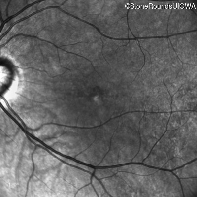
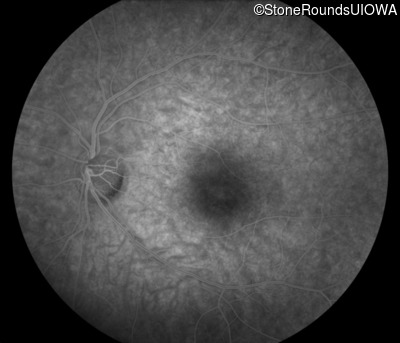
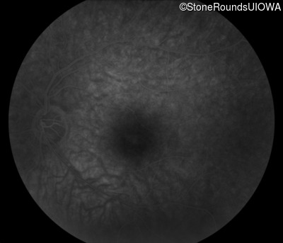
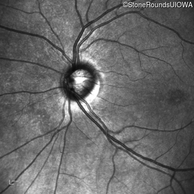
Infrared Fundus Photograph - Right - 20/32 -2
Exemplar
Infrared Fundus Photograph - Left - 20/80 -2
Exemplar
Visit at age: 42 years
Optical Coherence Tomography - Right - 20/63
Exemplar / OCT Stack
OCT Stack
OCT Stack
Optical Coherence Tomography - Left - 20/80
Exemplar / OCT Stack
OCT Stack
OCT Stack
Infrared Fundus Photograph - Right - 20/63
Exemplar

Infrared Fundus Photograph - Left - 20/80
Exemplar
Visit at age: 43 years
Optical Coherence Tomography - Right - 20/160 +1
Exemplar / OCT Stack
OCT Stack
OCT Stack
Optical Coherence Tomography - Left - 20/200 -1
Exemplar / OCT Stack

OCT Stack
OCT Stack
Infrared Fundus Photograph - Right - 20/160 +1
Exemplar
Infrared Fundus Photograph - Left - 20/200 -1
Exemplar
Visit at age: 43 years (Visit 2)
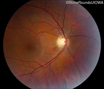
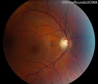
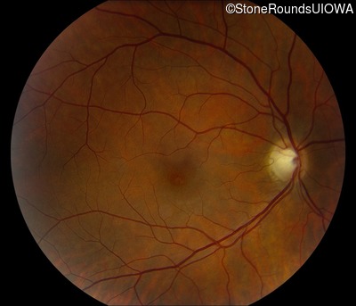
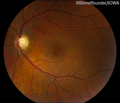
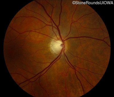
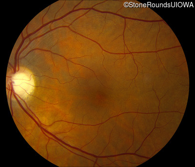
Fundus Photography - Right - 20/80 -1
Exemplar
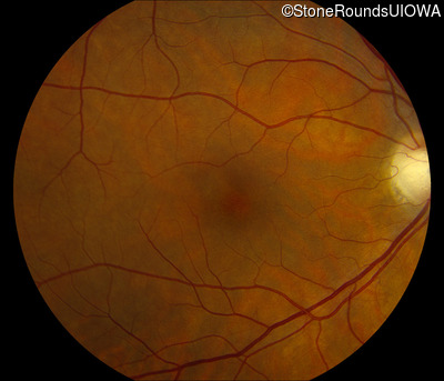
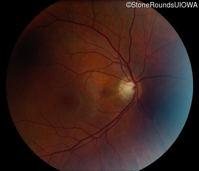
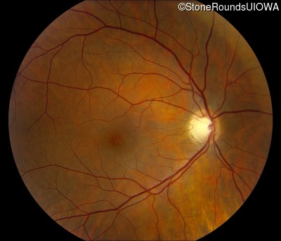
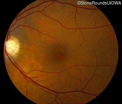
Fundus Photography - Left - 20/125
Exemplar
Goldmann Visual Field - Right - 20/80 -1
Exemplar
Goldmann Visual Field - Left - 20/125
Exemplar
Visit at age: 44 years
Optical Coherence Tomography - Right - 20/200
Exemplar / OCT Stack
OCT Stack
OCT Stack
Optical Coherence Tomography - Left - 20/200
Exemplar / OCT Stack
OCT Stack
OCT Stack
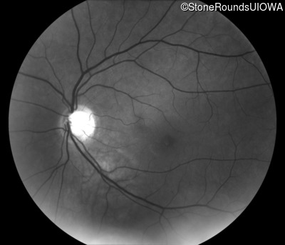
Infrared Fundus Photograph - Right - 20/200
Exemplar

Infrared Fundus Photograph - Left - 20/200
Exemplar
Visit at age: 46 years
Goldmann Visual Field - Right - 20/160 -1
Exemplar
Goldmann Visual Field - Left - 20/200 +1
Exemplar
Case Level Images
Diagnosis & molecular findings
| Disease | Gene | Allele 1 variant(s) | Allele 2 variant(s) | Inheritance mode |
|---|---|---|---|---|
| AR Cone and Cone Rod Dystrophy | MFSD8 | Glu336Gln GAG>CAG | IVS6+5 G>A | AR |
Disease:
Gene:
Allele 1:
Glu336Gln GAG>CAG
Allele 2:
IVS6+5 G>A
Inheritance:
AR