Case
SR1899
Student Mode
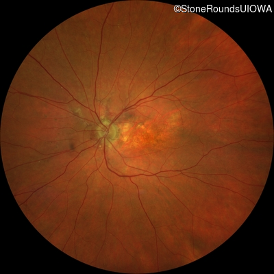
Pseudoxanthoma Elasticum (IID2)
Male
Male
Hidden
SR1899
Student Mode
Pseudoxanthoma Elasticum (IID2)
Male
Male
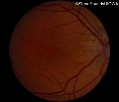
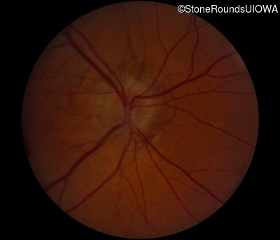
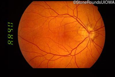
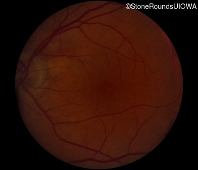
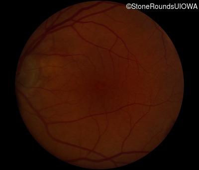
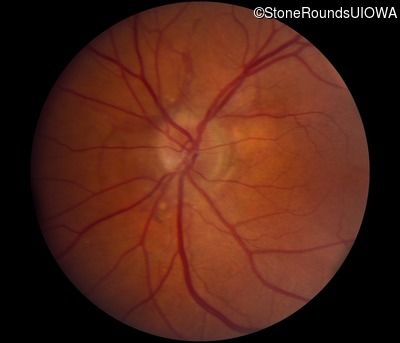
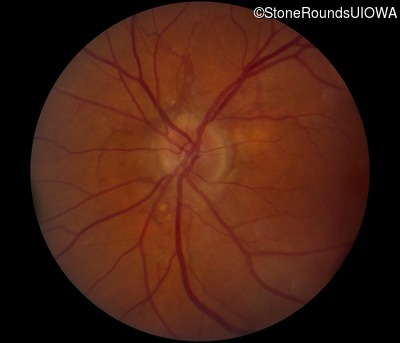
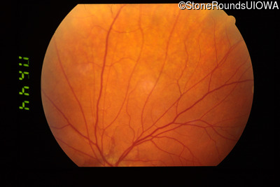
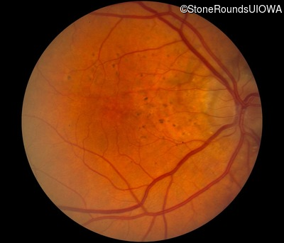
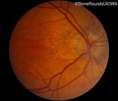
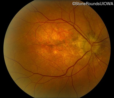
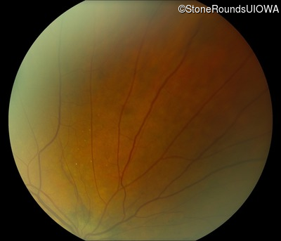
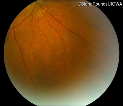
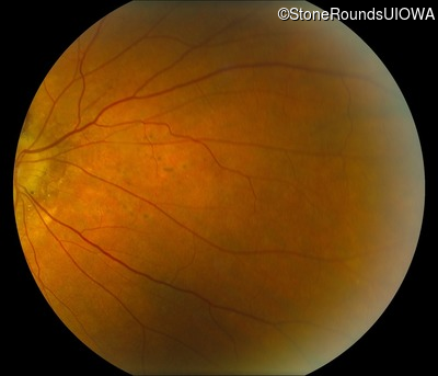
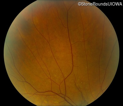
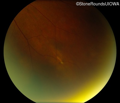
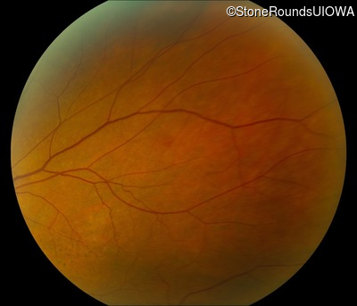
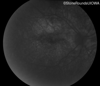
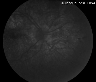
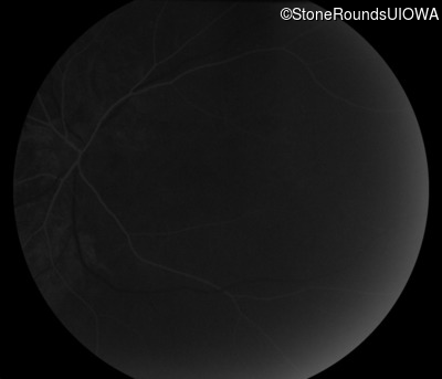
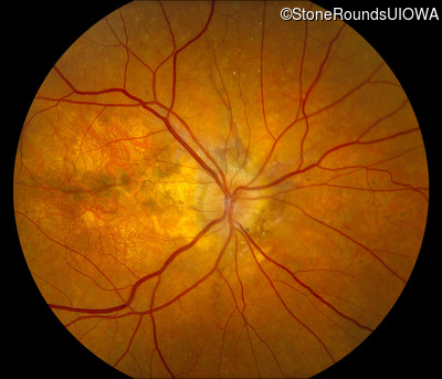
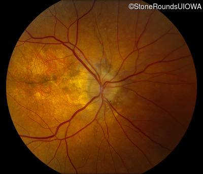
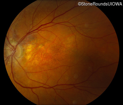
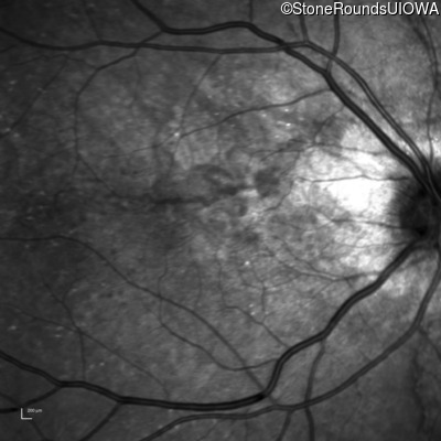
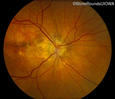
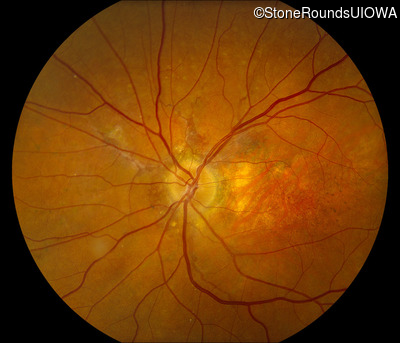
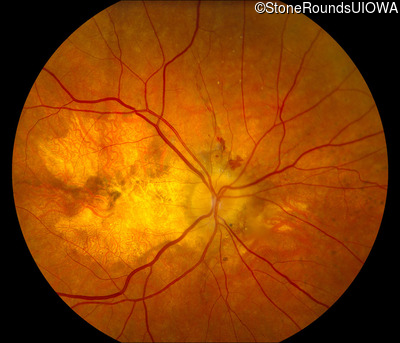
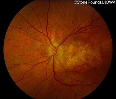
Visit at age: 49 years
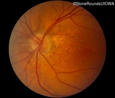
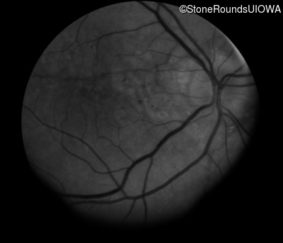
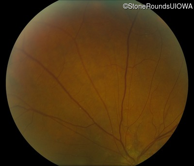
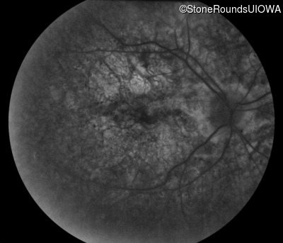
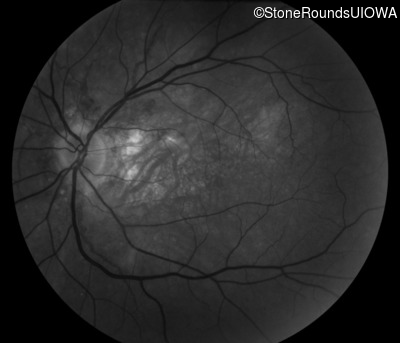
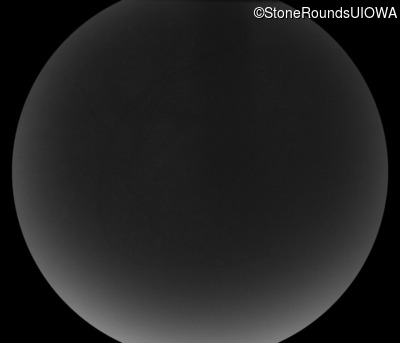
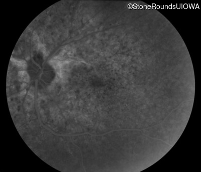
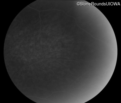
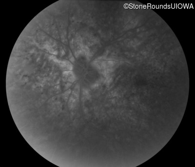
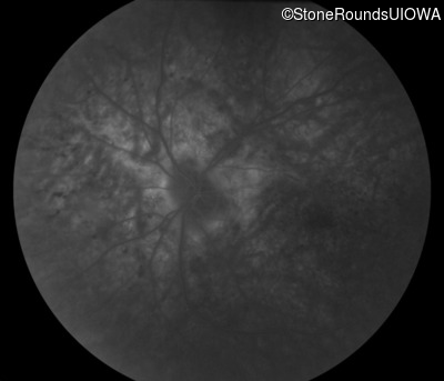
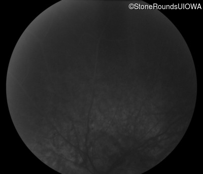
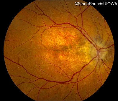
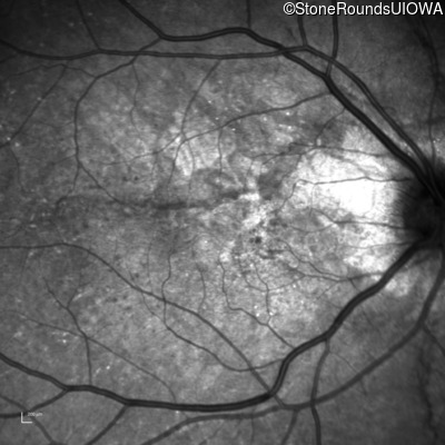
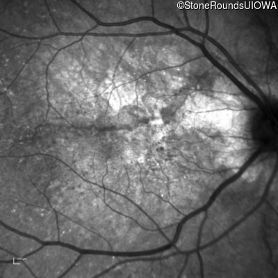
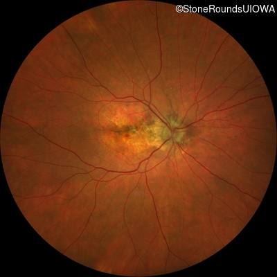
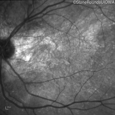
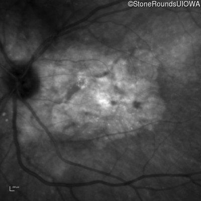
Fundus Photography - Right - 20/15 -2
Exemplar
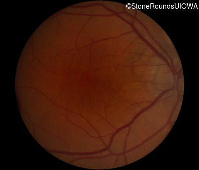
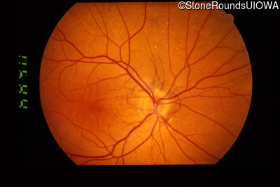
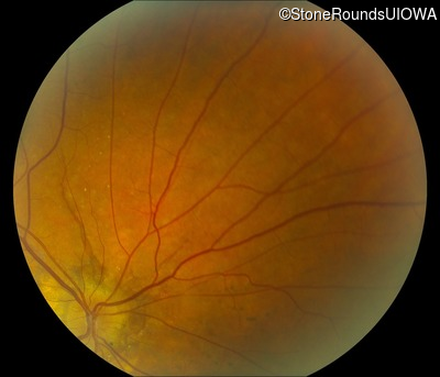
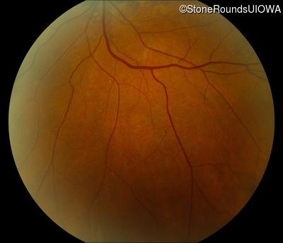
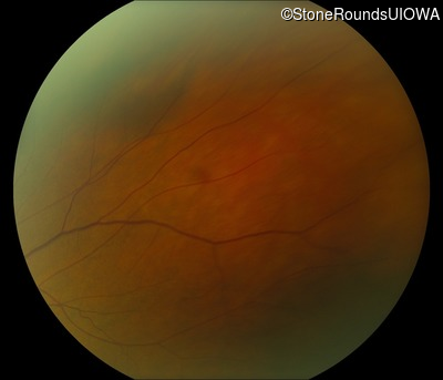
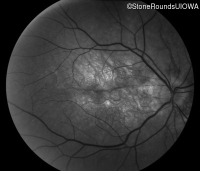
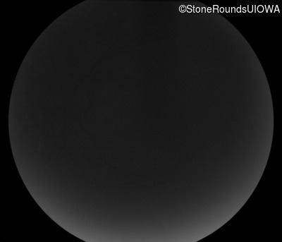
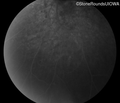
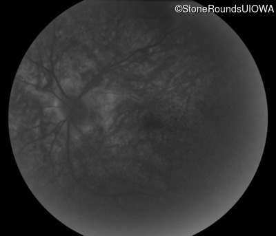
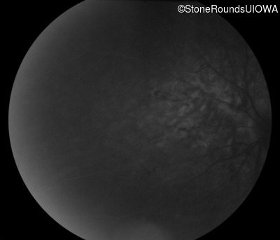
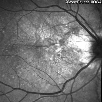
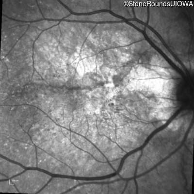
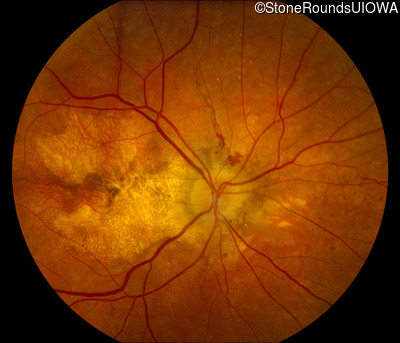
Fundus Photography - Left - 20/15 -2
Exemplar
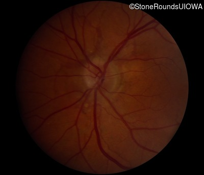
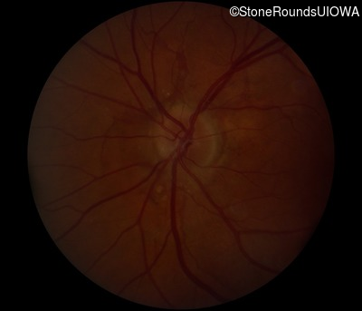
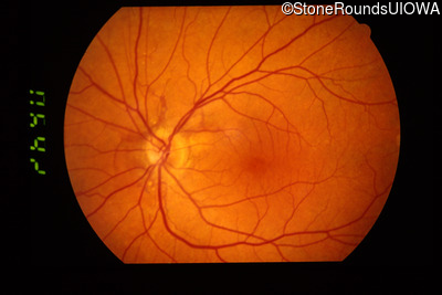
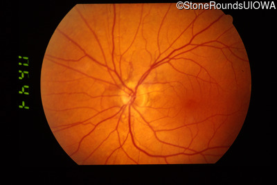
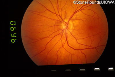
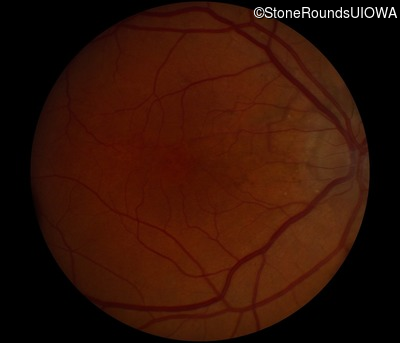
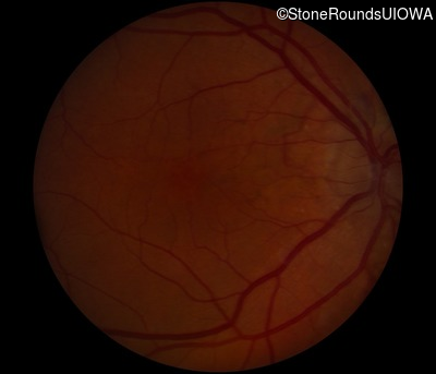
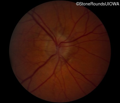
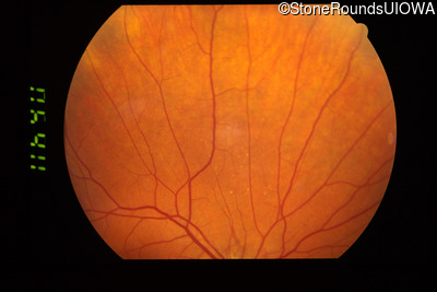
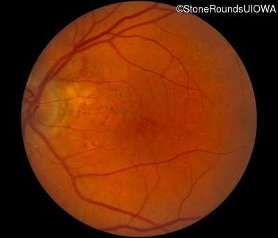
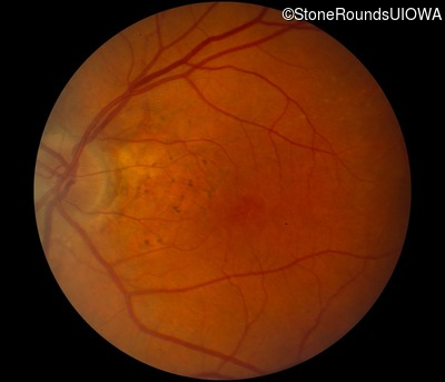
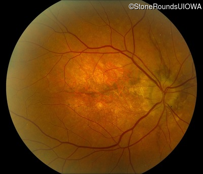
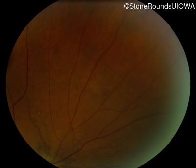
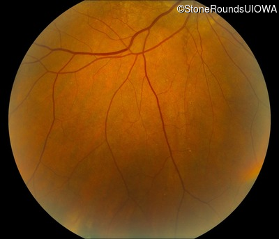
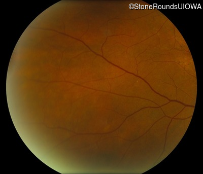
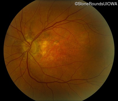
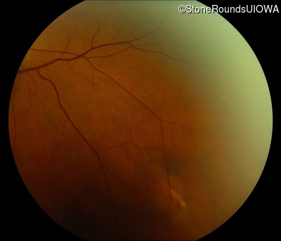
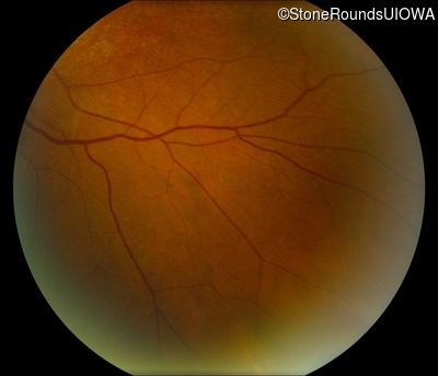
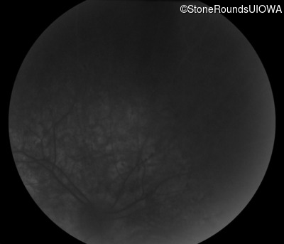
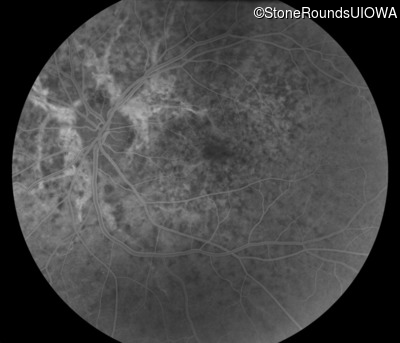
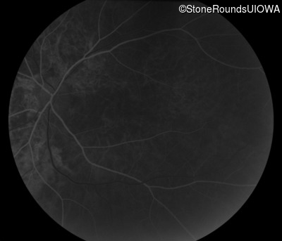
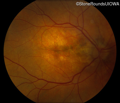
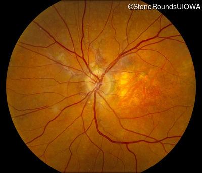
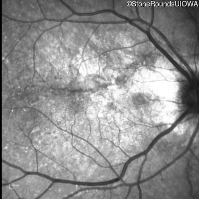
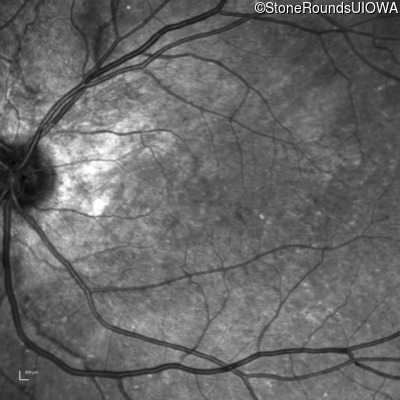
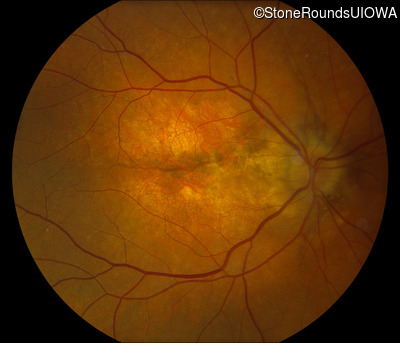
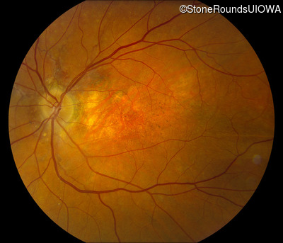
Visit at age: 51 years
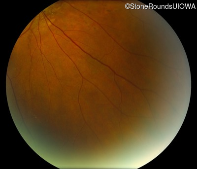
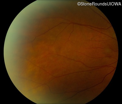
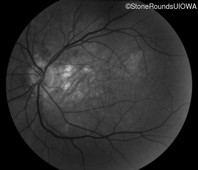
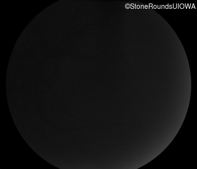
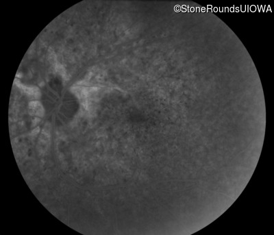
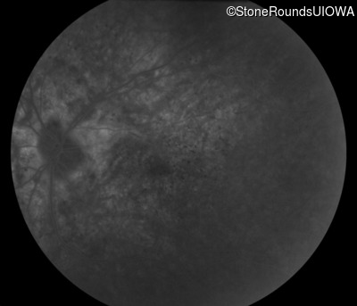
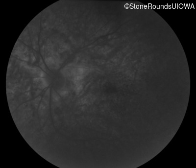
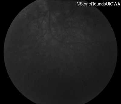
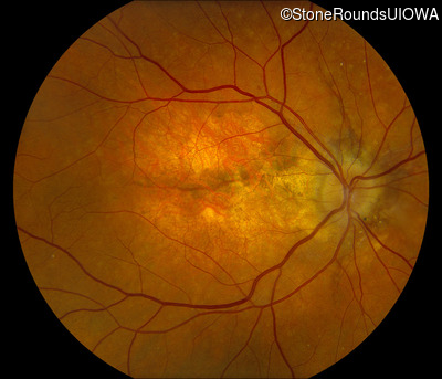
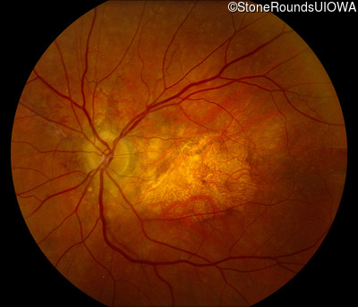
Fundus Photography - Right - 20/15 -2
Exemplar
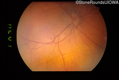
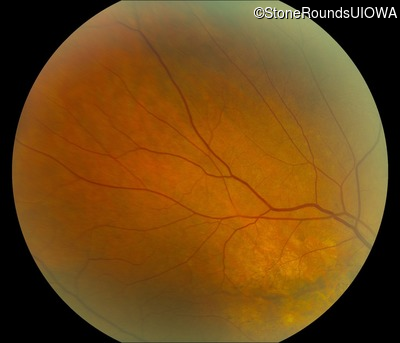
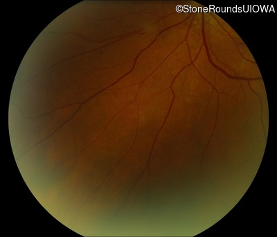
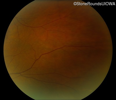
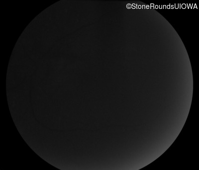
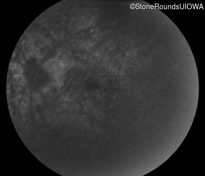
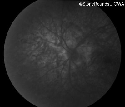
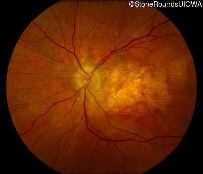
Fundus Photography - Left - 20/15 -2
Exemplar

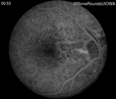
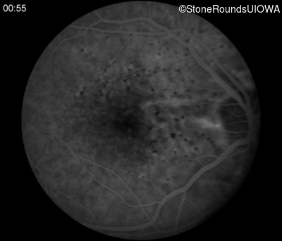
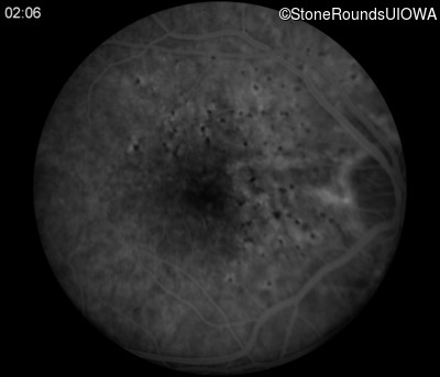
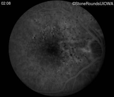
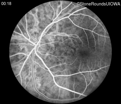
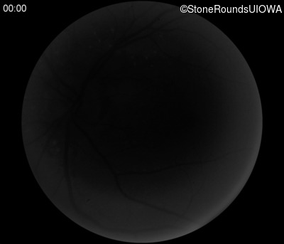
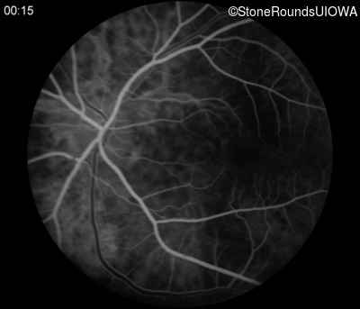
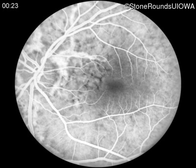
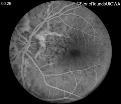
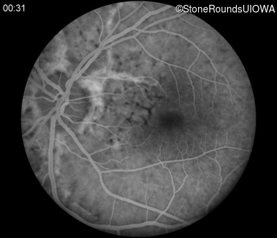
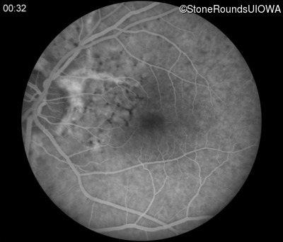
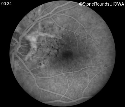
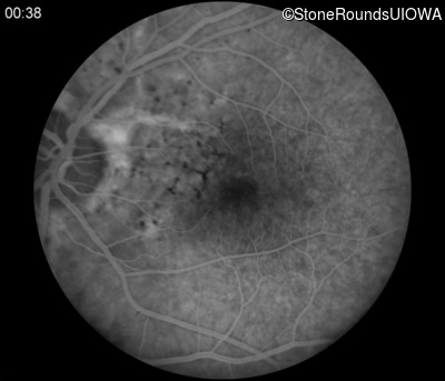
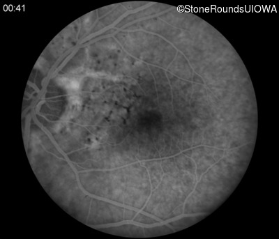
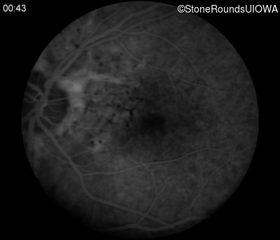
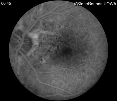
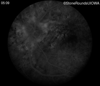
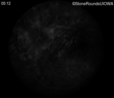
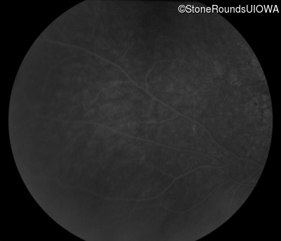
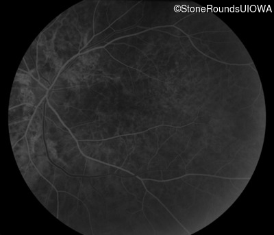
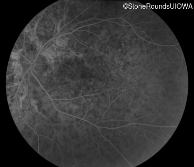
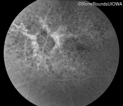
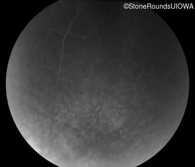
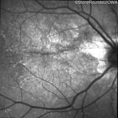
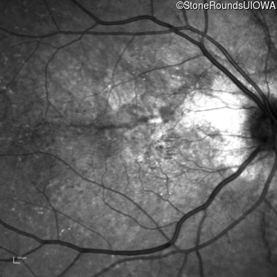
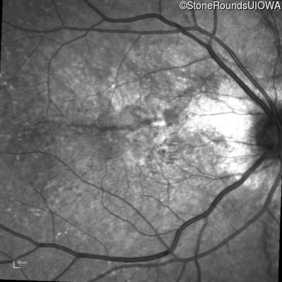
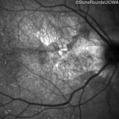
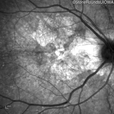
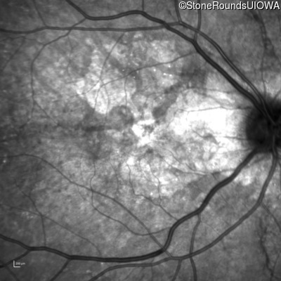
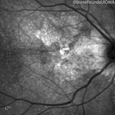
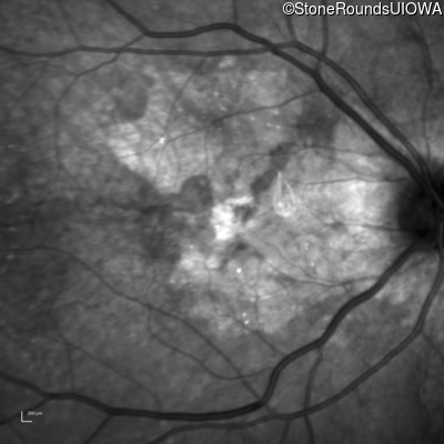
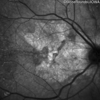
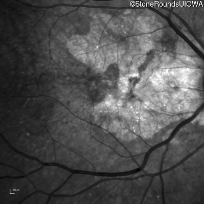
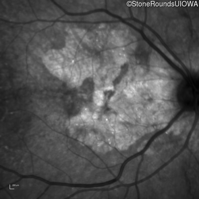
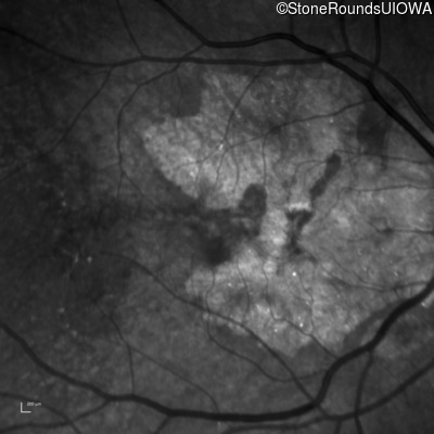
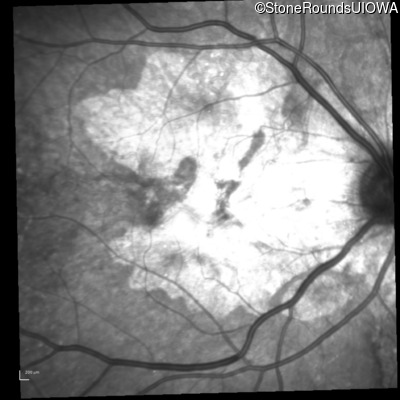
Fluorescein Angiography - Right - 20/15 -2
Exemplar

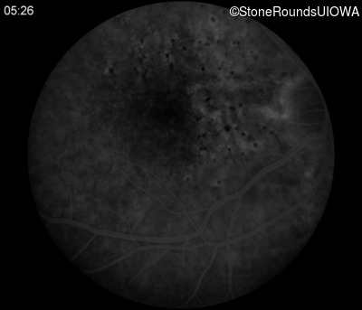
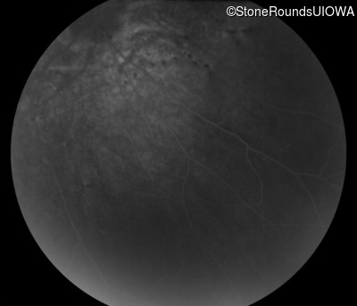
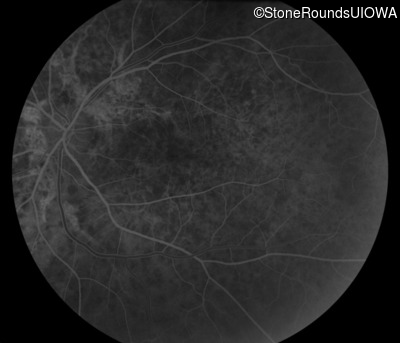
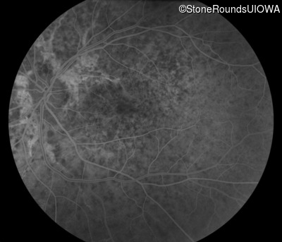
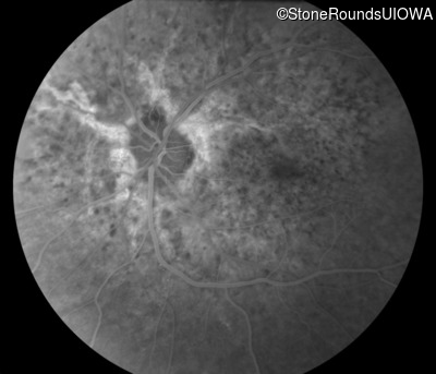
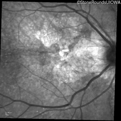
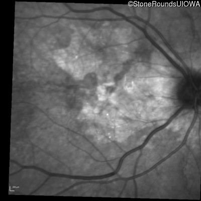
Fluorescein Angiography - Left - 20/15 -2
Exemplar


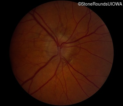
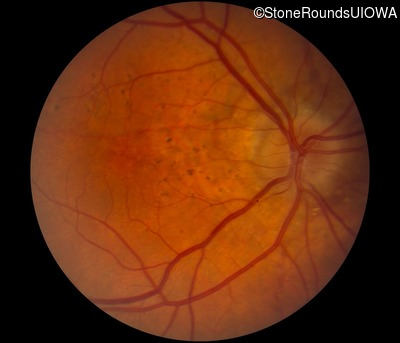
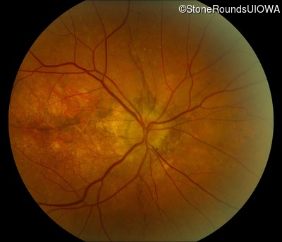
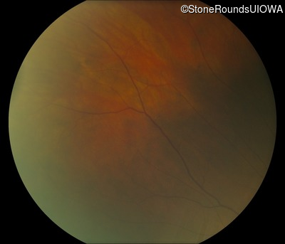
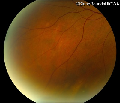
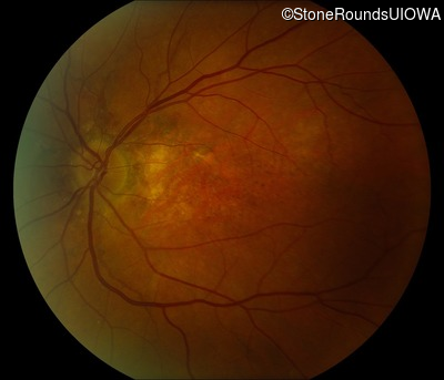
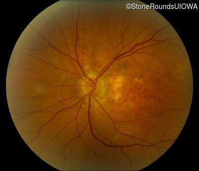
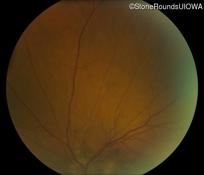
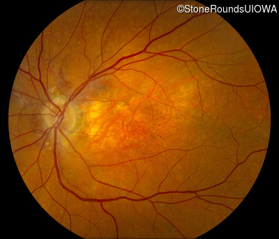
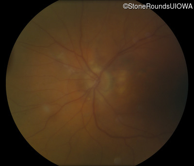
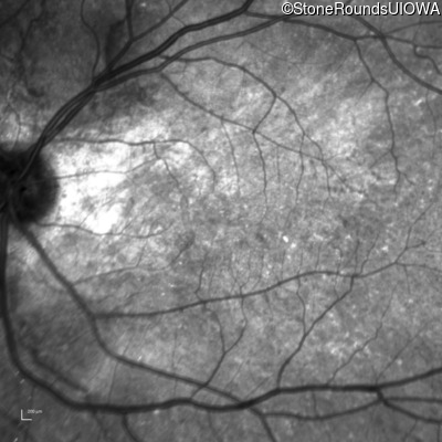
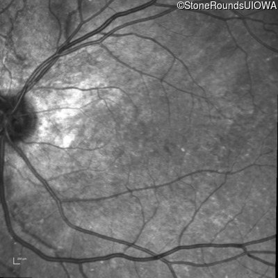
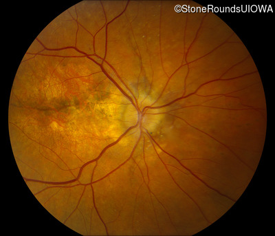
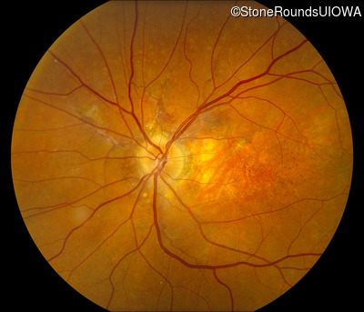
Visit at age: 61 years
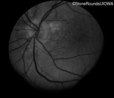
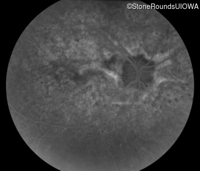
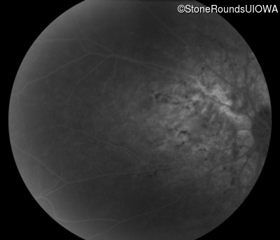
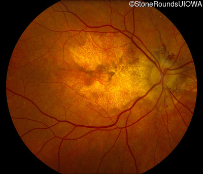
Fundus Photography - Right - 20/20 -1
Exemplar
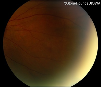

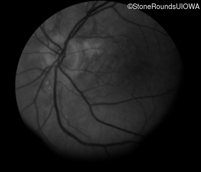
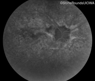
Fundus Photography - Left - 20/20 -2
Exemplar
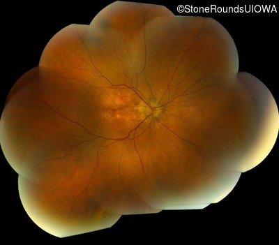
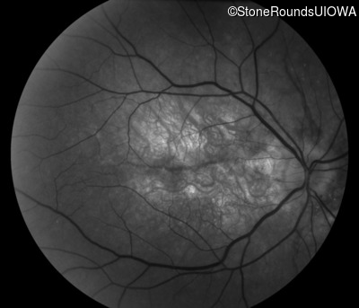
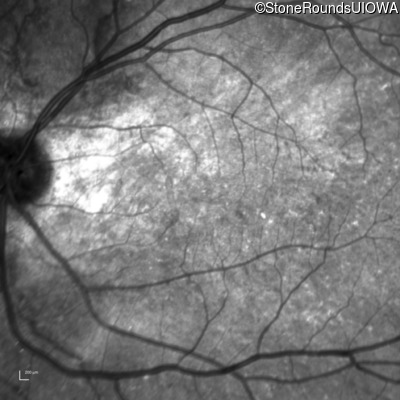
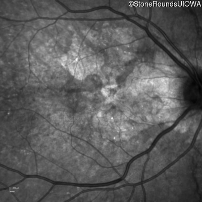
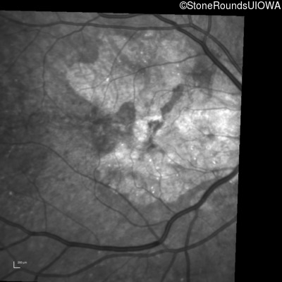
Fundus Montage - Right - 20/20 -1
Exemplar
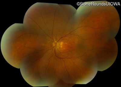
Fundus Montage - Left - 20/20 -2
Exemplar
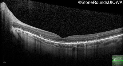
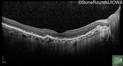
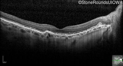
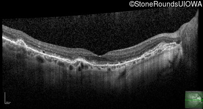
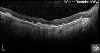
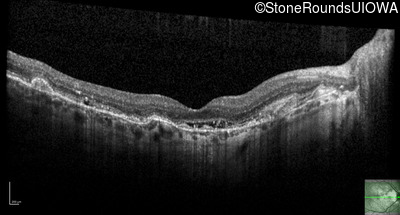
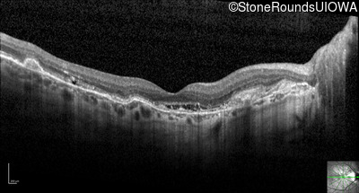
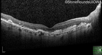
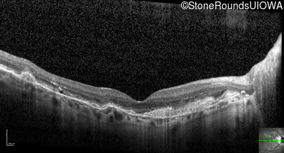
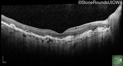
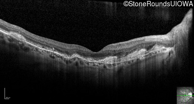
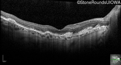
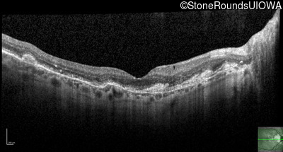
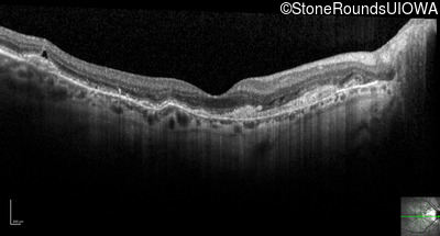
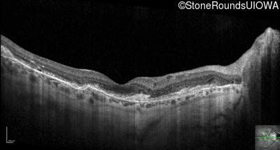
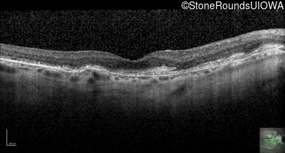
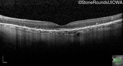
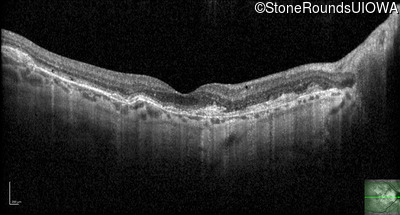
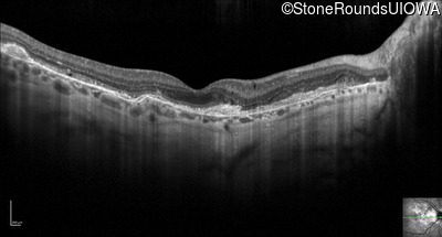
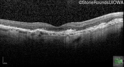
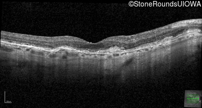
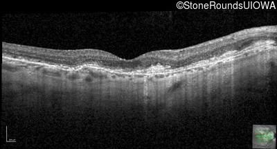
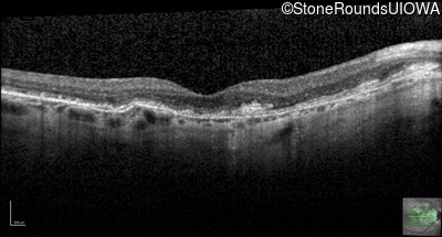
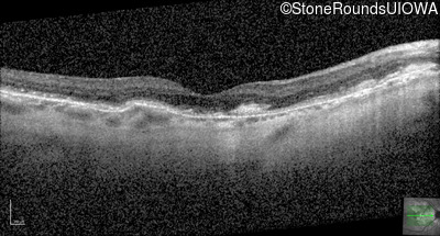
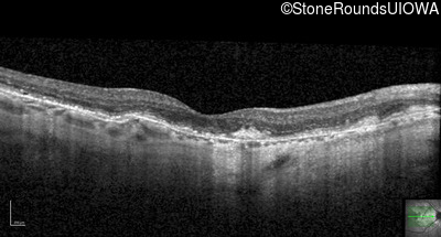
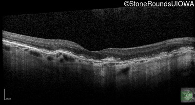
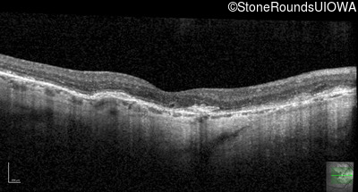
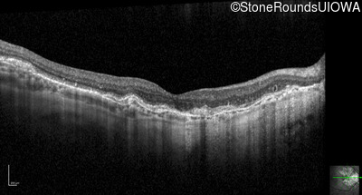
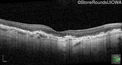
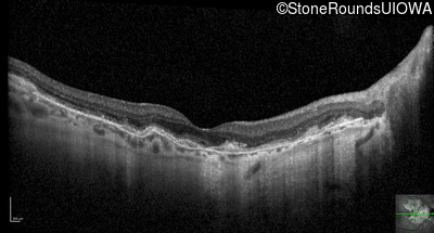
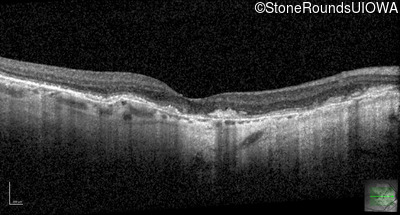
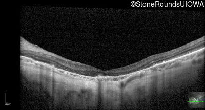
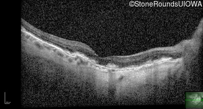
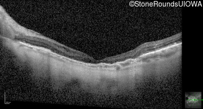
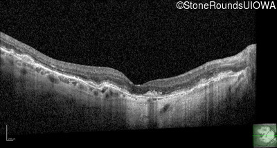
Optical Coherence Tomography - Right - 20/20 -1
Exemplar / OCT Stack
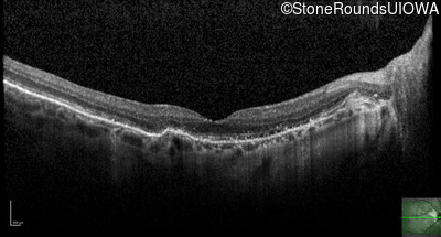
OCT Stack
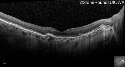
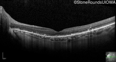
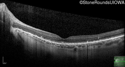
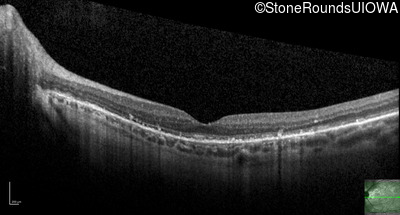
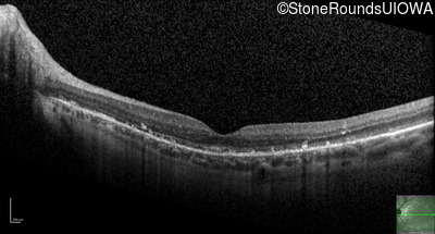
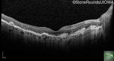
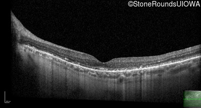
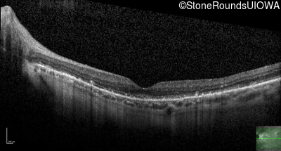
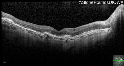
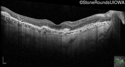
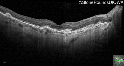
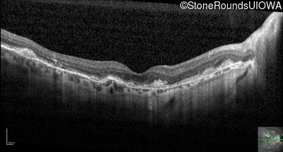
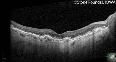
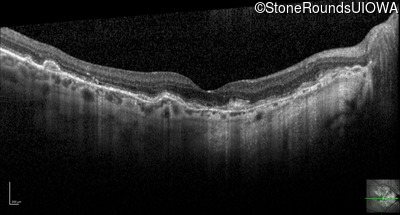
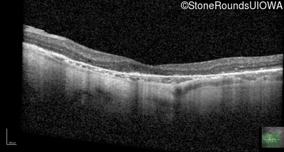
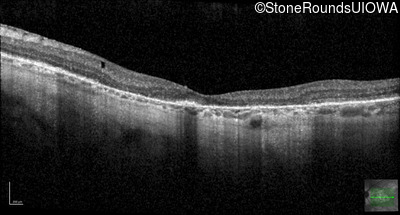
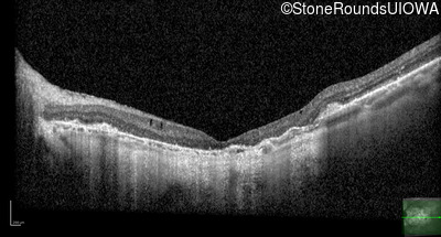
Optical Coherence Tomography - Left - 20/20 -2
Exemplar / OCT Stack
OCT Stack
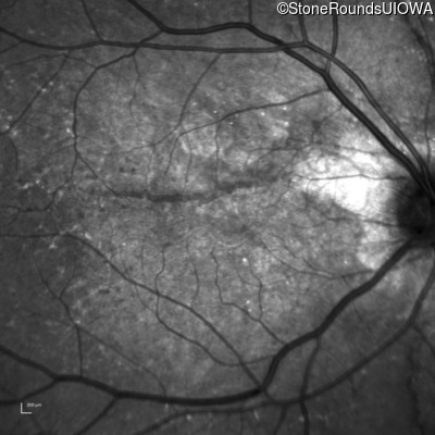
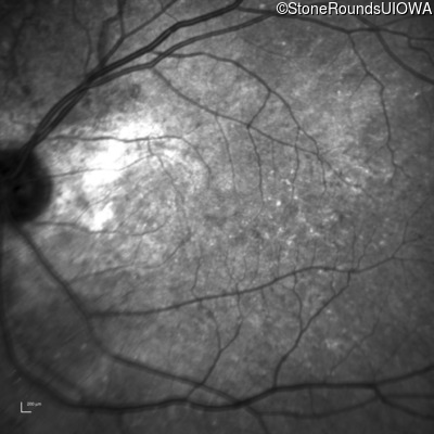
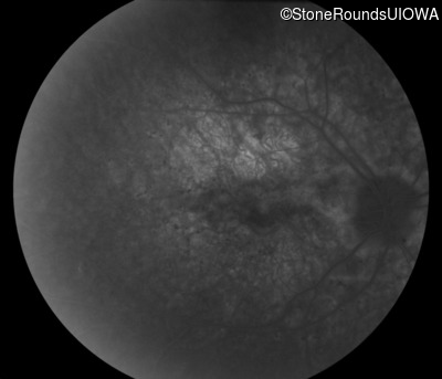
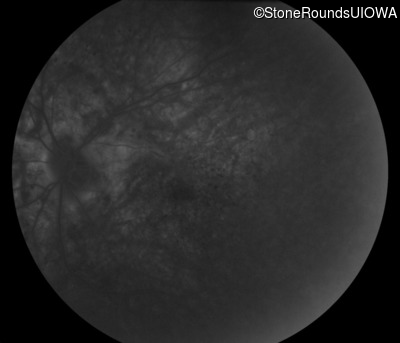
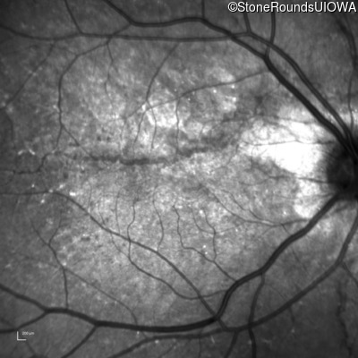
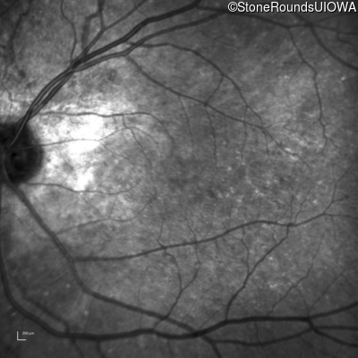
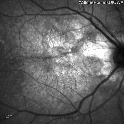
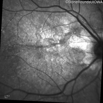
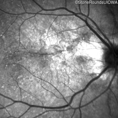
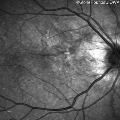
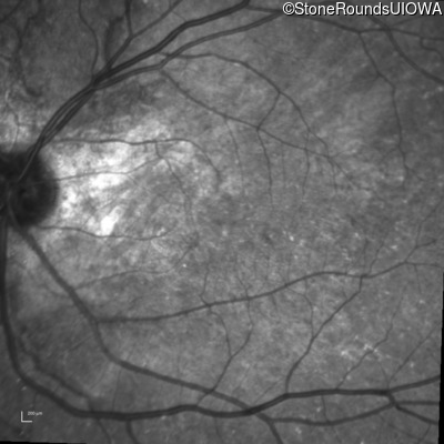
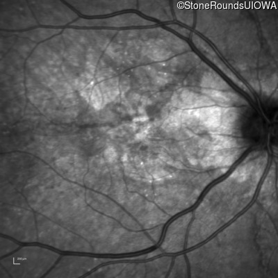
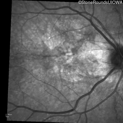
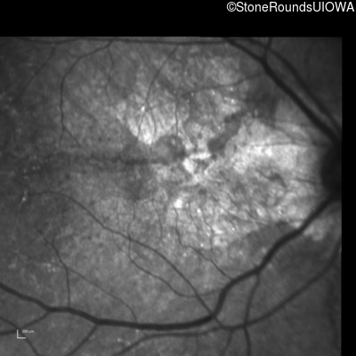
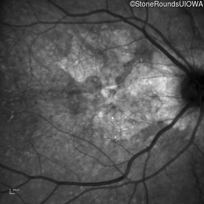
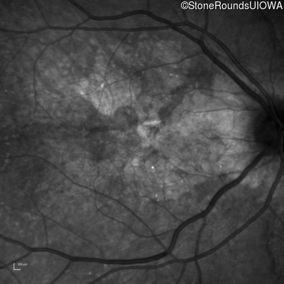
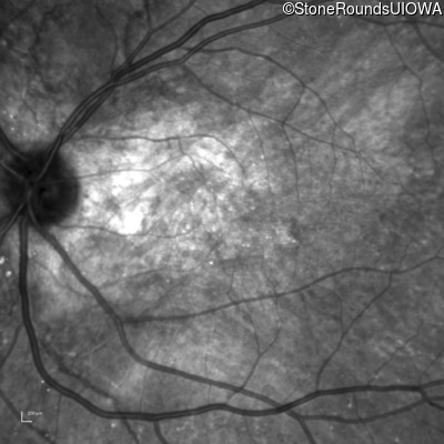
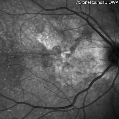
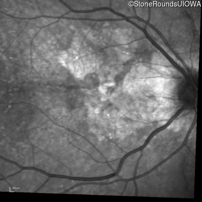
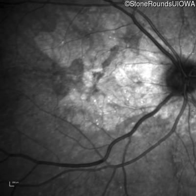
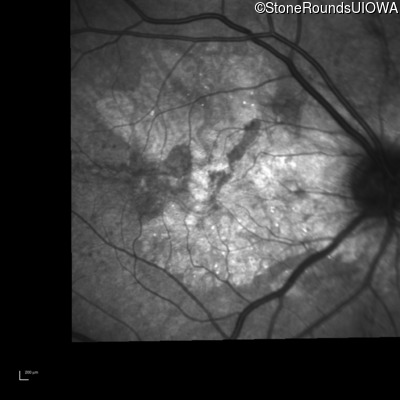
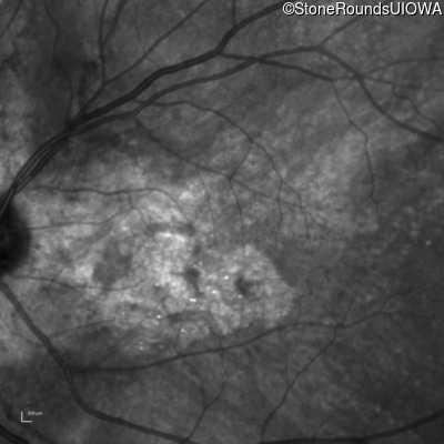
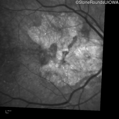
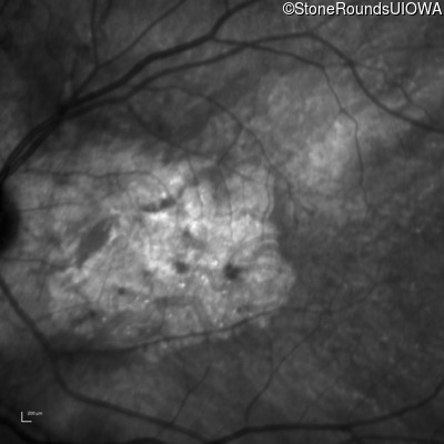
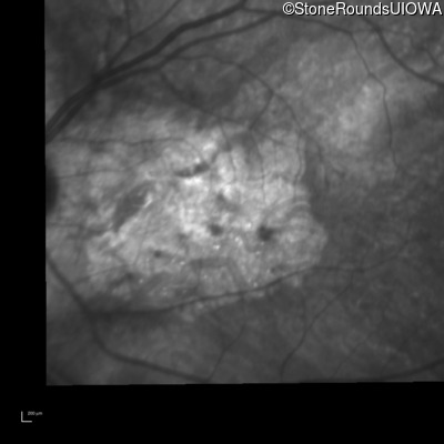
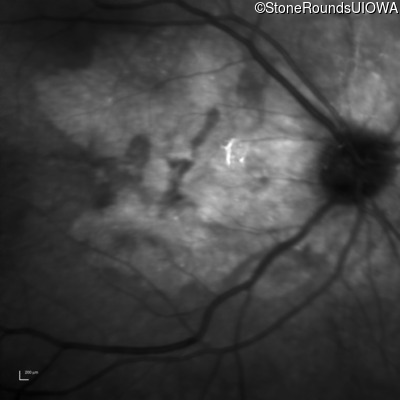
Infrared Fundus Photograph - Right - 20/20 -1
Exemplar

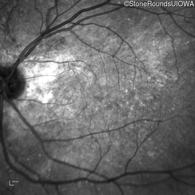
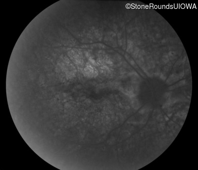
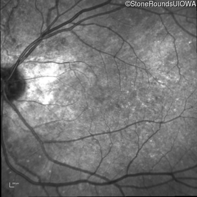
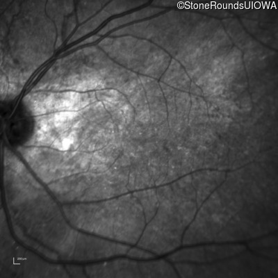
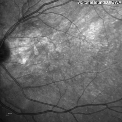
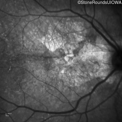
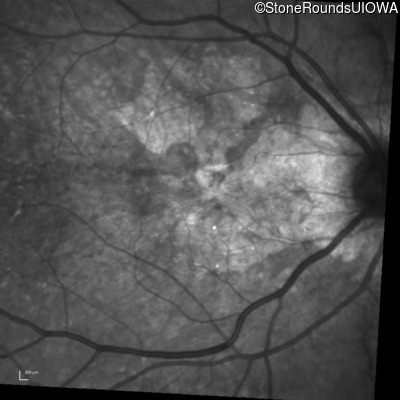
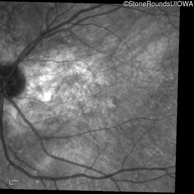
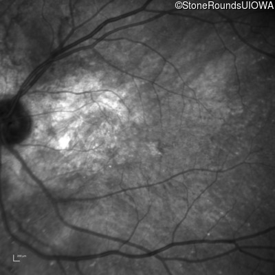
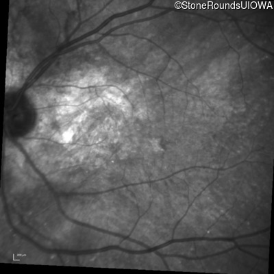
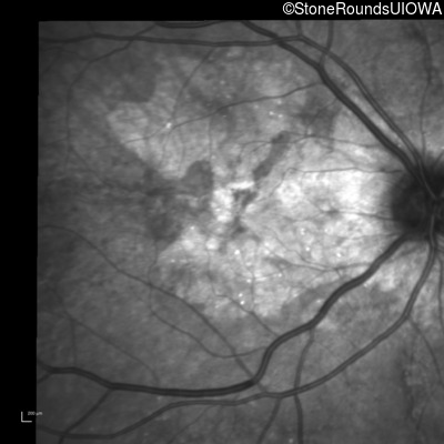
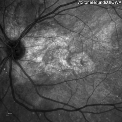
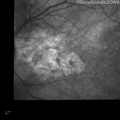
Infrared Fundus Photograph - Left - 20/20 -2
Exemplar
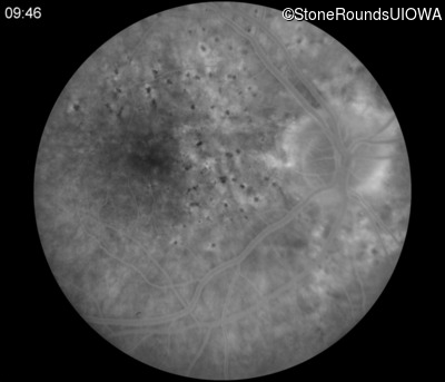
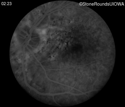
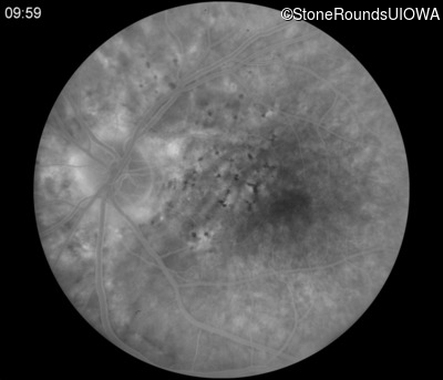
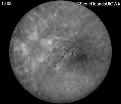
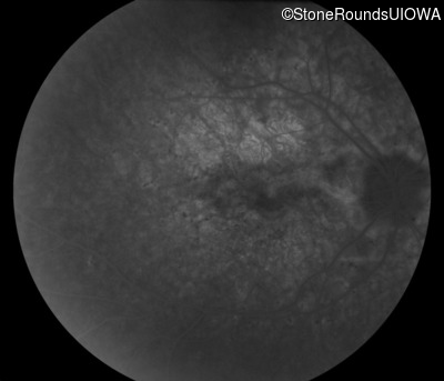
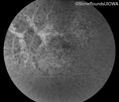
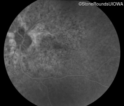
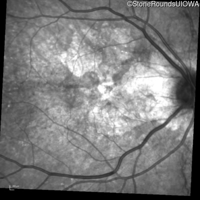
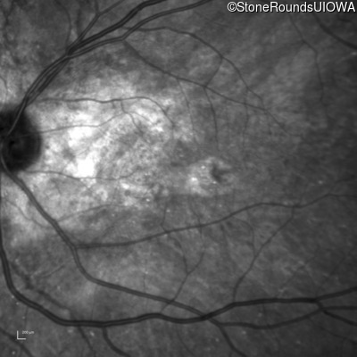
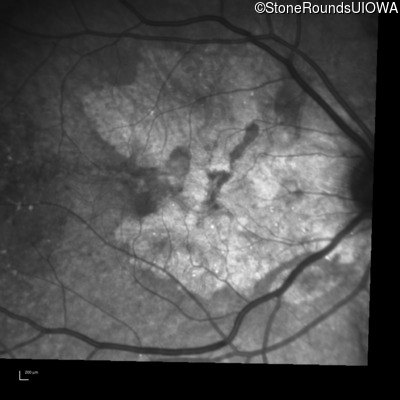
Fluorescein Angiography - Right - 20/20 -1
Exemplar

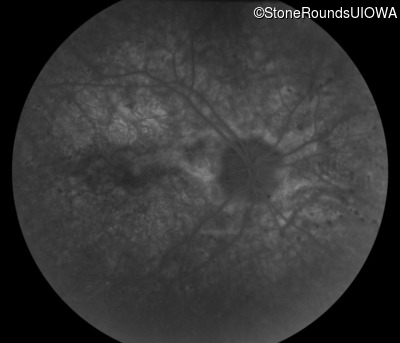
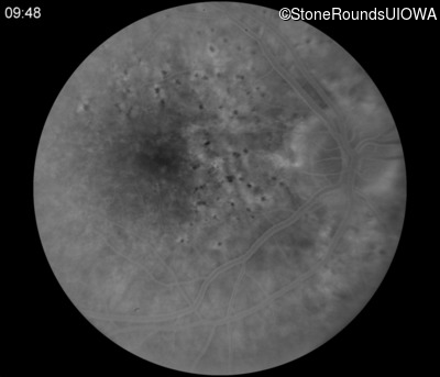
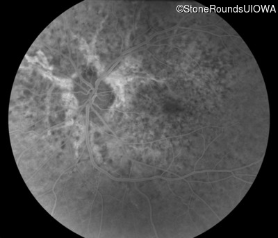
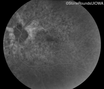
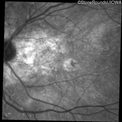
Fluorescein Angiography - Left - 20/20 -2
Exemplar


Visit at age: 61 years (Visit 2)
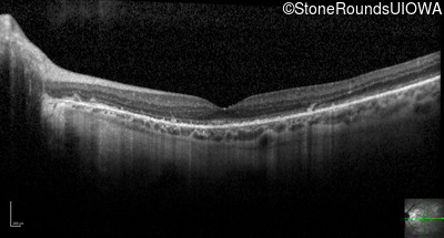
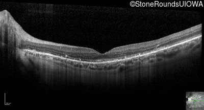
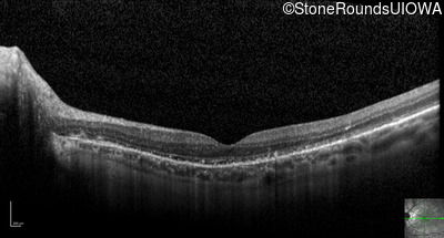
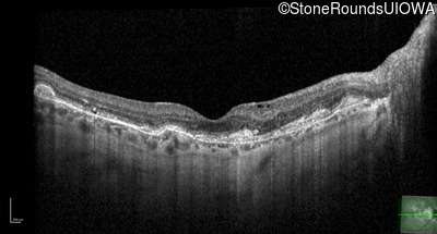
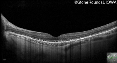
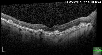
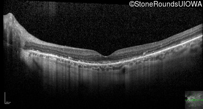
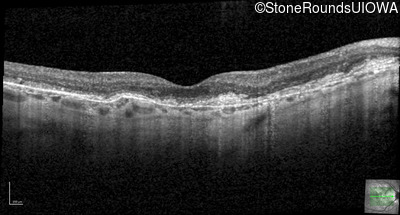
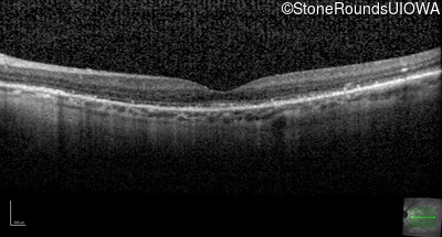
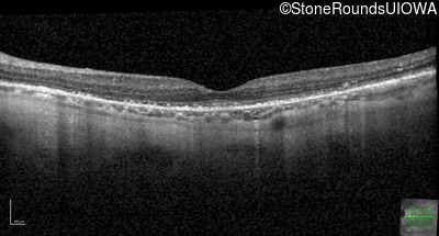
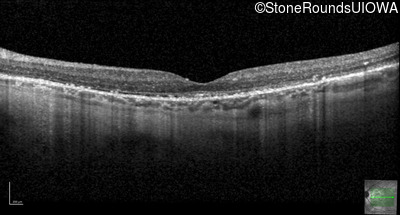
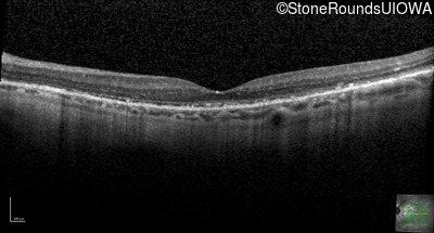
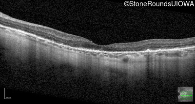
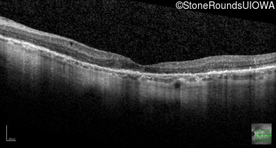
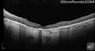
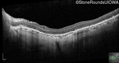
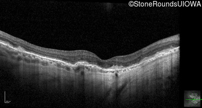
Optical Coherence Tomography - Right - 20/25 +1
Exemplar / OCT Stack
OCT Stack
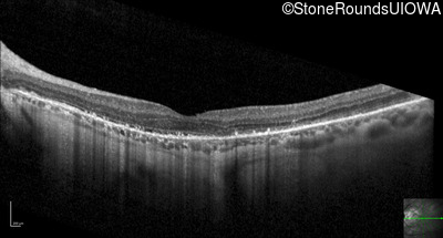
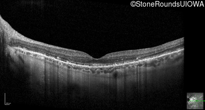
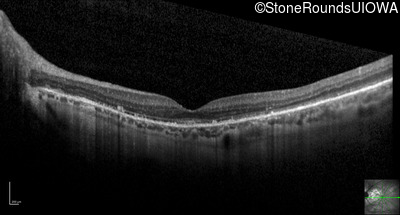
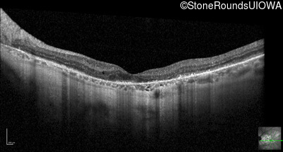
Optical Coherence Tomography - Left - 20/25 +3
Exemplar / OCT Stack
OCT Stack
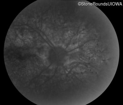
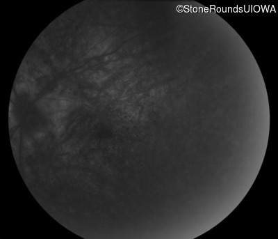
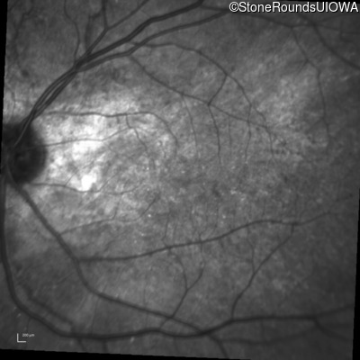
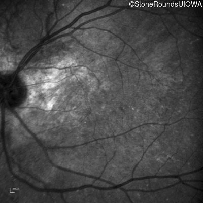
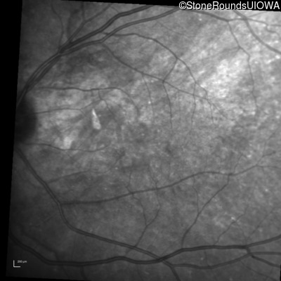
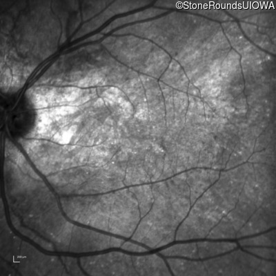
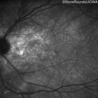
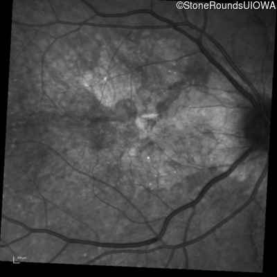
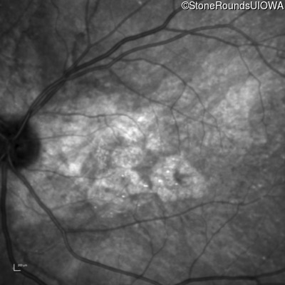
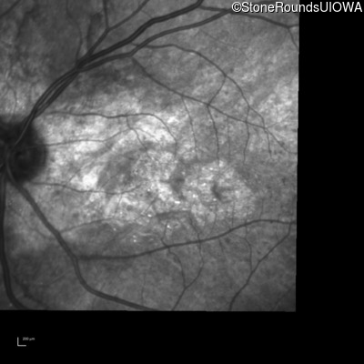
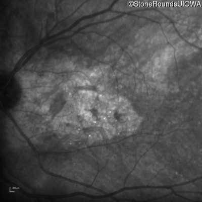
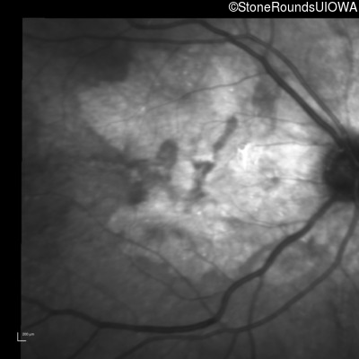
Infrared Fundus Photograph - Right - 20/25 +1
Exemplar
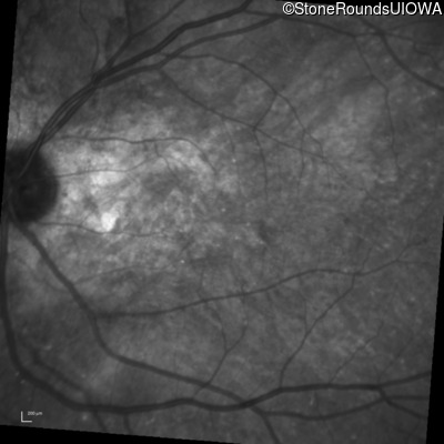
Infrared Fundus Photograph - Left - 20/25 +3
Exemplar
Visit at age: 62 years
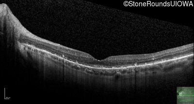
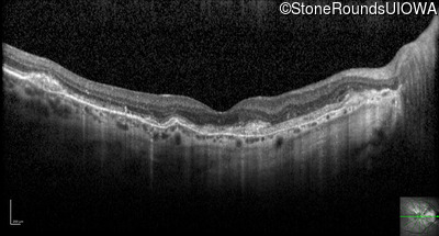
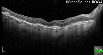
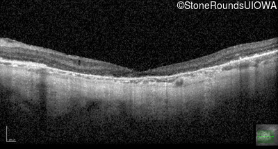
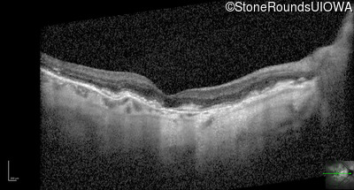
Optical Coherence Tomography - Right - 20/20 -2
Exemplar / OCT Stack
OCT Stack
Optical Coherence Tomography - Left - 20/20 -3
Exemplar / OCT Stack
OCT Stack
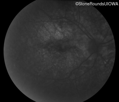
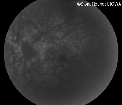
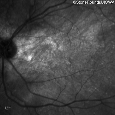
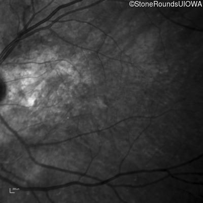
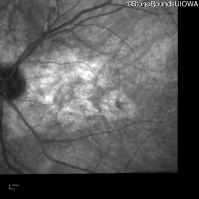
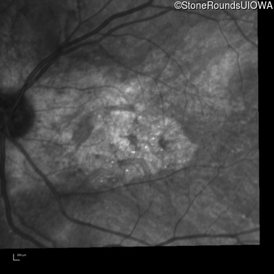
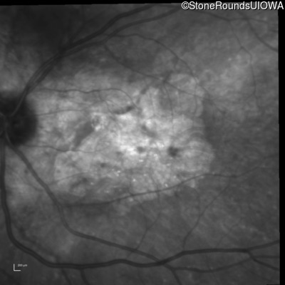
Infrared Fundus Photograph - Right - 20/20 -2
Exemplar
Infrared Fundus Photograph - Left - 20/20 -3
Exemplar
Visit at age: 62 years (Visit 2)
Fundus Photography - Right - 20/50 +1
Exemplar
Fundus Photography - Left - 20/25 -2
Exemplar
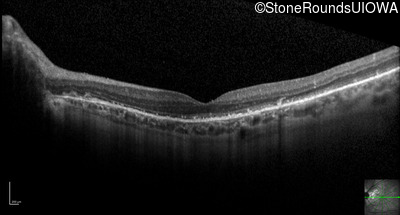
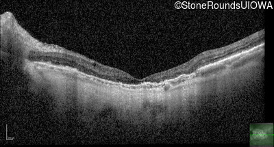
Optical Coherence Tomography - Right - 20/50 +1
Exemplar / OCT Stack
OCT Stack
Optical Coherence Tomography - Left - 20/25 -2
Exemplar / OCT Stack
OCT Stack
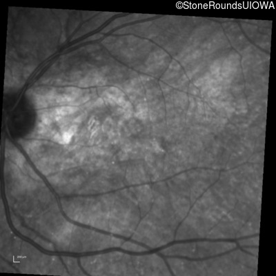
Infrared Fundus Photograph - Right - 20/50 +1
Exemplar
Infrared Fundus Photograph - Left - 20/25 -2
Exemplar
Visit at age: 62 years (Visit 3)
Optical Coherence Tomography - Right - 20/80
Exemplar / OCT Stack
OCT Stack
Optical Coherence Tomography - Left - 20/20 -1
Exemplar / OCT Stack
OCT Stack
Infrared Fundus Photograph - Right - 20/80
Exemplar
Infrared Fundus Photograph - Left - 20/20 -1
Exemplar
Visit at age: 63 years
Optical Coherence Tomography - Right - 20/50 -1
Exemplar / OCT Stack
OCT Stack
Infrared Fundus Photograph - Right - 20/50 -1
Exemplar
Visit at age: 63 years (Visit 2)
Optical Coherence Tomography - Right - 20/50 +2
Exemplar / OCT Stack
OCT Stack
Infrared Fundus Photograph - Right - 20/50 +2
Exemplar
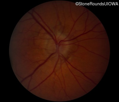
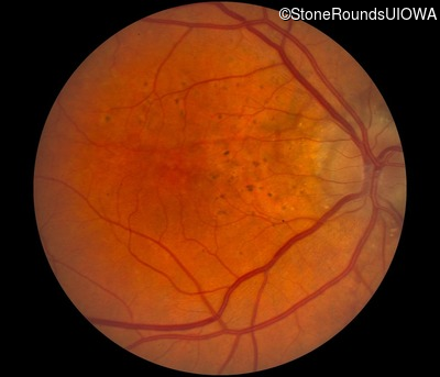
Visit at age: 63 years (Visit 3)
Fundus Photography - Right - 20/40 -2
Exemplar
Fundus Photography - Left - 20/20 -2
Exemplar
Optical Coherence Tomography - Right - 20/40 -2
Exemplar / OCT Stack
OCT Stack
Optical Coherence Tomography - Left - 20/20 -2
Exemplar / OCT Stack
OCT Stack
Infrared Fundus Photograph - Right - 20/40 -2
Exemplar
Infrared Fundus Photograph - Left - 20/20 -2
Exemplar
Visit at age: 63 years (Visit 4)
Optical Coherence Tomography - Right - 20/30 -2
Exemplar / OCT Stack
OCT Stack
Optical Coherence Tomography - Left - 20/25 -1
Exemplar / OCT Stack
OCT Stack
Infrared Fundus Photograph - Right - 20/30 -2
Exemplar
Infrared Fundus Photograph - Left - 20/25 -1
Exemplar
Visit at age: 63 years (Visit 5)
Fundus Photography - Right - 20/125
Exemplar
Fundus Photography - Left - 20/25
Exemplar
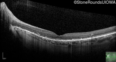
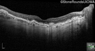
Optical Coherence Tomography - Right - 20/125
Exemplar / OCT Stack
OCT Stack
OCT Stack
Optical Coherence Tomography - Left - 20/25
Exemplar / OCT Stack
OCT Stack
OCT Stack
Infrared Fundus Photograph - Right - 20/125
Exemplar
Infrared Fundus Photograph - Left - 20/25
Exemplar
Visit at age: 64 years
Optical Coherence Tomography - Right - 20/60 -2
Exemplar / OCT Stack
OCT Stack
OCT Stack
Infrared Fundus Photograph - Right - 20/60 -2
Exemplar
Visit at age: 64 years (Visit 2)
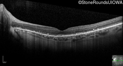
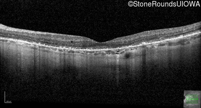
Optical Coherence Tomography - Right - 20/100
Exemplar / OCT Stack
OCT Stack
Optical Coherence Tomography - Left - 20/25 +1
Exemplar / OCT Stack
OCT Stack

Infrared Fundus Photograph - Right - 20/100
Exemplar
Infrared Fundus Photograph - Left - 20/25 +1
Exemplar
Visit at age: 64 years (Visit 3)
Optical Coherence Tomography - Right - 20/60 -2
Exemplar / OCT Stack
OCT Stack
Optical Coherence Tomography - Left - 20/20 -3
Exemplar / OCT Stack
OCT Stack
Infrared Fundus Photograph - Right - 20/60 -2
Exemplar
Infrared Fundus Photograph - Left - 20/20 -3
Exemplar
Visit at age: 64 years (Visit 4)
Optical Coherence Tomography - Right - 20/100
Exemplar / OCT Stack
OCT Stack
Optical Coherence Tomography - Left - 20/20 -1
Exemplar / OCT Stack
OCT Stack
Infrared Fundus Photograph - Right - 20/100
Exemplar
Infrared Fundus Photograph - Left - 20/20 -1
Exemplar
Visit at age: 64 years (Visit 5)
Optical Coherence Tomography - Right - 20/100 +1
Exemplar / OCT Stack
OCT Stack
Optical Coherence Tomography - Left - 20/25
Exemplar / OCT Stack
OCT Stack
Infrared Fundus Photograph - Right - 20/100 +1
Exemplar
Infrared Fundus Photograph - Left - 20/25
Exemplar
Visit at age: 64 years (Visit 6)
Optical Coherence Tomography - Right - 20/40
Exemplar / OCT Stack
OCT Stack
Optical Coherence Tomography - Left - 20/20 -2
Exemplar / OCT Stack
OCT Stack
Infrared Fundus Photograph - Right - 20/40
Exemplar
Infrared Fundus Photograph - Left - 20/20 -2
Exemplar
Visit at age: 65 years
Optical Coherence Tomography - Right - 20/80
Exemplar / OCT Stack
OCT Stack
Optical Coherence Tomography - Left - 20/30
Exemplar / OCT Stack
OCT Stack
Infrared Fundus Photograph - Right - 20/80
Exemplar
Infrared Fundus Photograph - Left - 20/30
Exemplar
Visit at age: 65 years (Visit 2)
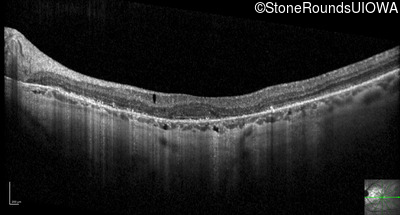
Optical Coherence Tomography - Right - 20/150
Exemplar / OCT Stack
OCT Stack
Optical Coherence Tomography - Left - 20/40 -2
Exemplar / OCT Stack
OCT Stack

Infrared Fundus Photograph - Right - 20/150
Exemplar
Infrared Fundus Photograph - Left - 20/40 -2
Exemplar
Visit at age: 65 years (Visit 3)
Optical Coherence Tomography - Right - 20/150
Exemplar / OCT Stack
OCT Stack
Optical Coherence Tomography - Left - 20/30
Exemplar / OCT Stack
OCT Stack
Infrared Fundus Photograph - Right - 20/150
Exemplar
Infrared Fundus Photograph - Left - 20/30
Exemplar
Visit at age: 65 years (Visit 4)
Optical Coherence Tomography - Right - 20/200 -2
Exemplar / OCT Stack
OCT Stack
Optical Coherence Tomography - Left - 20/63
Exemplar / OCT Stack
OCT Stack
Infrared Fundus Photograph - Right - 20/200 -2
Exemplar
Infrared Fundus Photograph - Left - 20/63
Exemplar
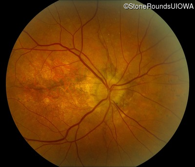
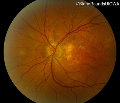
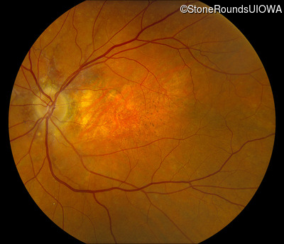
Visit at age: 66 years
Fundus Photography - Right - 20/125
Exemplar
Fundus Photography - Left - 20/125
Exemplar
Optical Coherence Tomography - Right - 20/125
Exemplar / OCT Stack
OCT Stack
Optical Coherence Tomography - Left - 20/125
Exemplar / OCT Stack
OCT Stack
Infrared Fundus Photograph - Right - 20/125
Exemplar
Infrared Fundus Photograph - Left - 20/125
Exemplar
Visit at age: 67 years
Optical Coherence Tomography - Right - 20/200 -3
Exemplar / OCT Stack
OCT Stack
Optical Coherence Tomography - Left - 20/200 -3
Exemplar / OCT Stack
OCT Stack
Infrared Fundus Photograph - Right - 20/200 -3
Exemplar
Infrared Fundus Photograph - Left - 20/200 -3
Exemplar
Visit at age: 68 years
Optical Coherence Tomography - Right - 20/150
Exemplar / OCT Stack
Optical Coherence Tomography - Left - 20/150
Exemplar / OCT Stack
Infrared Fundus Photograph - Right - 20/150
Exemplar
Infrared Fundus Photograph - Left - 20/150
Exemplar
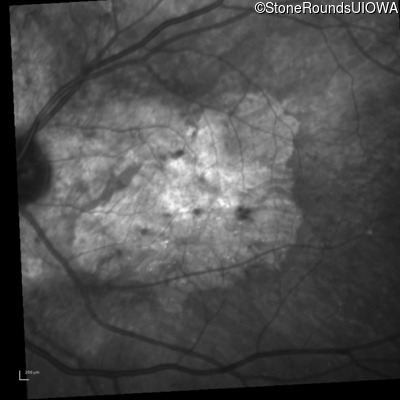
Case Level Images
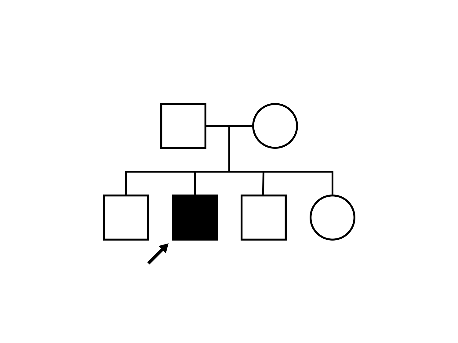
Diagnosis & molecular findings
| Disease | Gene | Allele 1 variant(s) | Allele 2 variant(s) | Inheritance mode |
|---|---|---|---|---|
| Pseudoxanthoma Elasticum | ABCC6 | Arg1141Stop CGA>TGA | Arg1398Stop CGA>TGA | AR |
Disease:
Gene:
Allele 1:
Arg1141Stop CGA>TGA
Allele 2:
Arg1398Stop CGA>TGA
Inheritance:
AR