Case
SR83
Student Mode
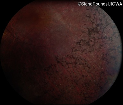
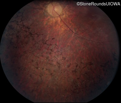
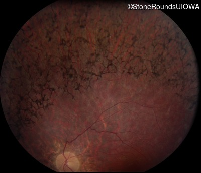
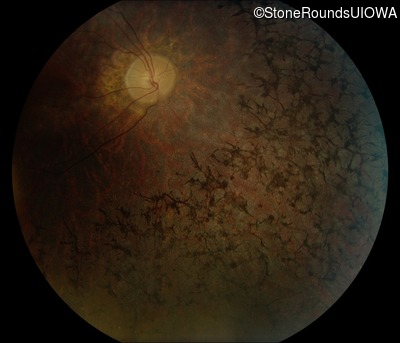
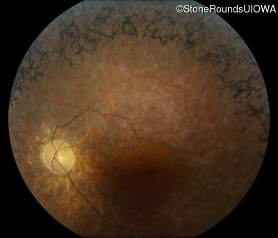
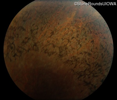
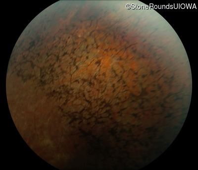
Posterior Column Ataxia with Retinitis Pigmentosa (IB7a)
Male
Male
Hidden
SR83
Student Mode
Posterior Column Ataxia with Retinitis Pigmentosa (IB7a)
Male
Male
Please Login or Register to download images or create a PowerPoint slideshow.
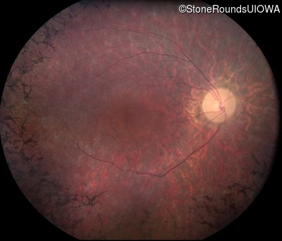
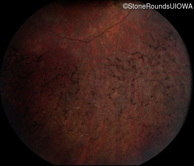
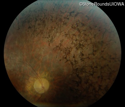
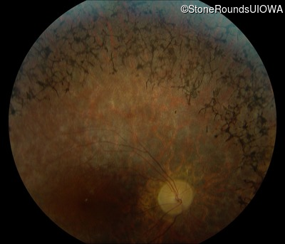
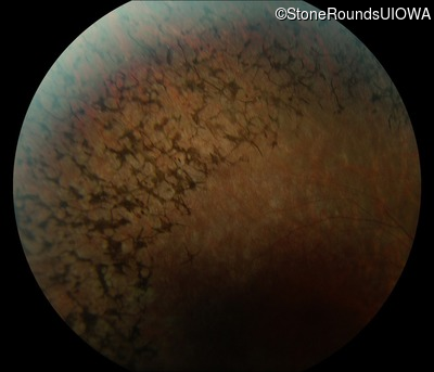
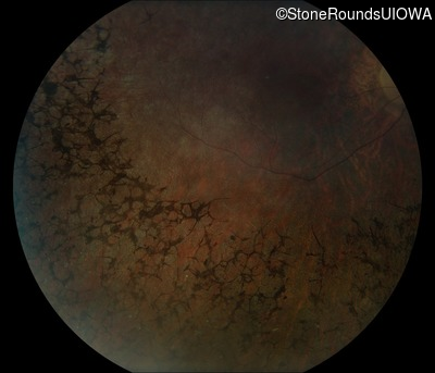
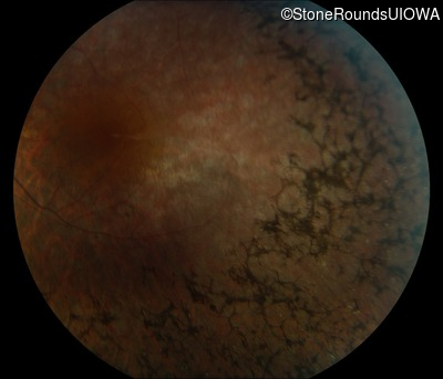
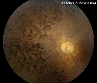
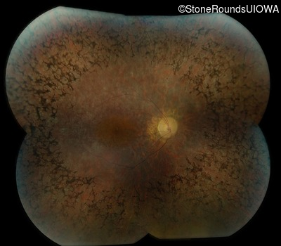
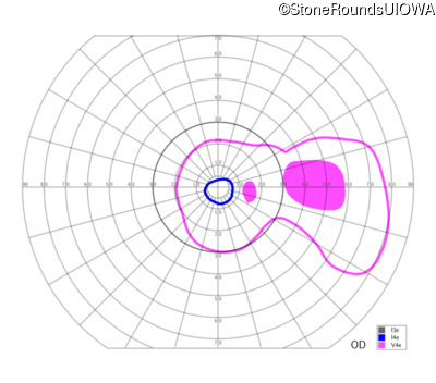
Visit at age: 21 years
Fundus Photography - Right - 20/40 sc
Exemplar
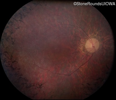
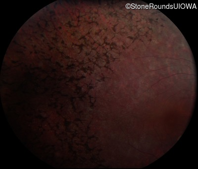
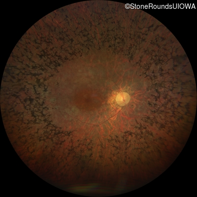
Fundus Photography - Left - 20/40 sc
Exemplar
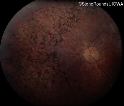
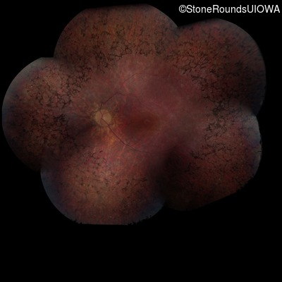
Fundus Montage - Right - 20/40 sc
Exemplar

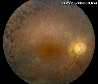
Fundus Montage - Left - 20/40 sc
Exemplar
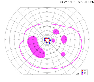
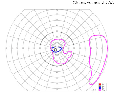
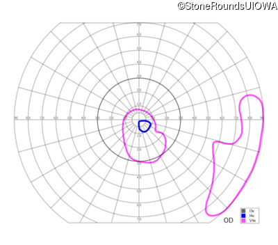
Goldmann Visual Field - Right - 20/40 sc
Exemplar
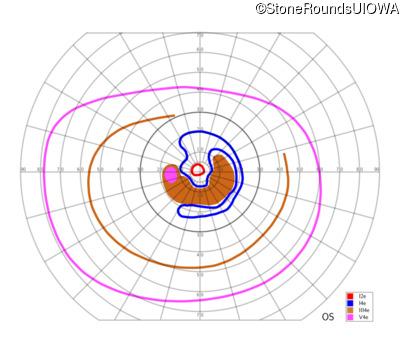
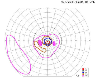
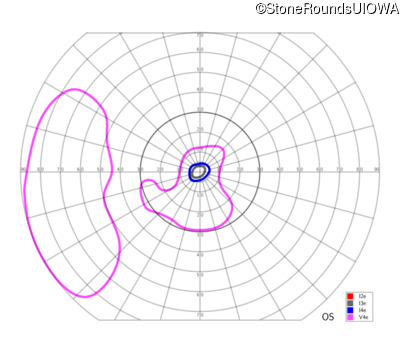
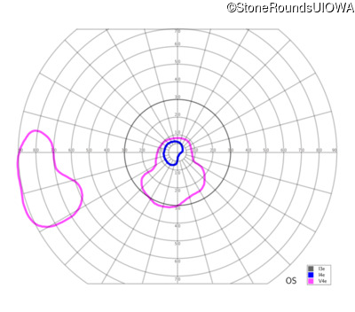
Goldmann Visual Field - Left - 20/40 sc
Exemplar
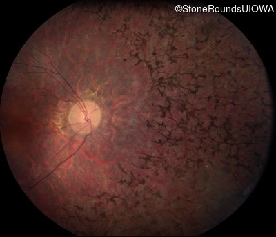
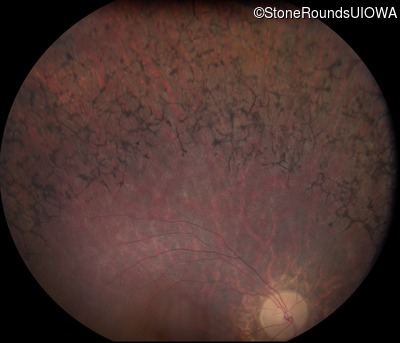
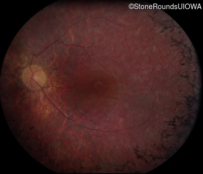
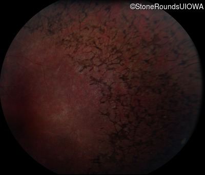
Visit at age: 28 years
Fundus Photography - Right - 20/60 +1 sc
Exemplar

Fundus Photography - Left - 20/50 -1 sc
Exemplar

Fundus Montage - Right - 20/60 +1 sc
Exemplar
Fundus Montage - Left - 20/50 -1 sc
Exemplar

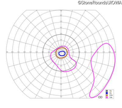
Goldmann Visual Field - Right - 20/60 +1 sc
Exemplar
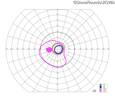
Goldmann Visual Field - Left - 20/50 -1 sc
Exemplar
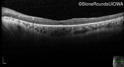
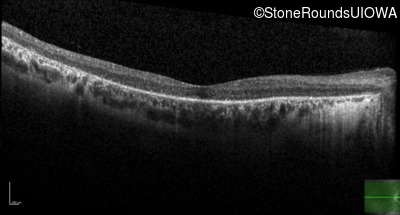
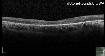
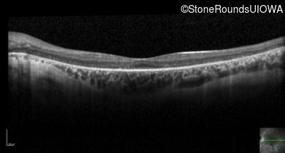
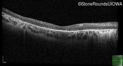
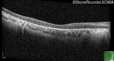
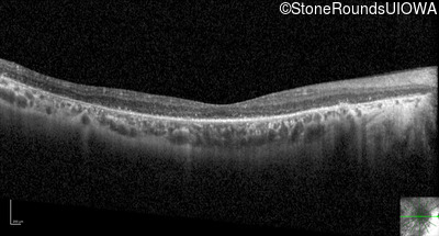
Optical Coherence Tomography - Right - 20/60 +1 sc
Exemplar / OCT Stack
OCT Stack
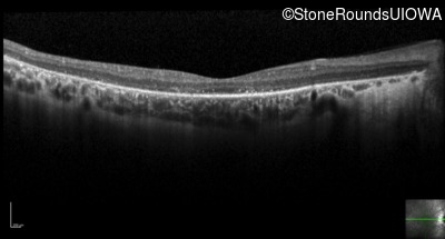
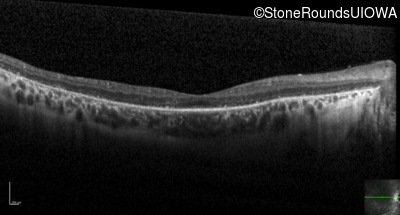
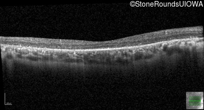
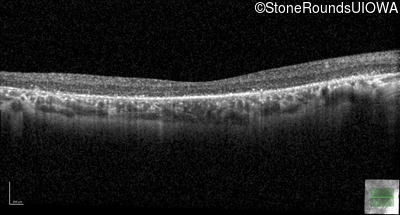
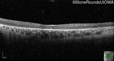
Optical Coherence Tomography - Left - 20/50 -1 sc
Exemplar / OCT Stack
OCT Stack
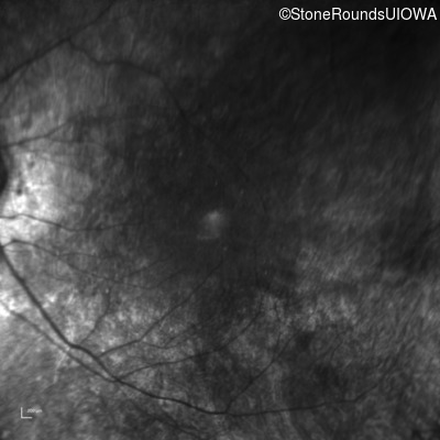
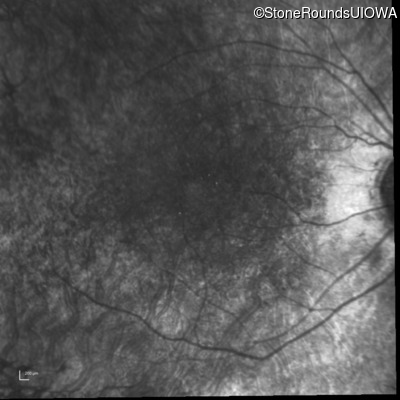
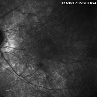
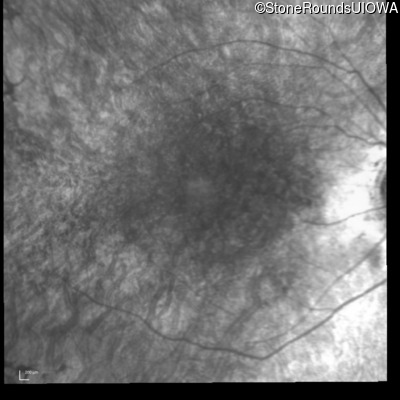
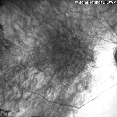
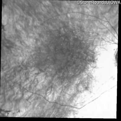
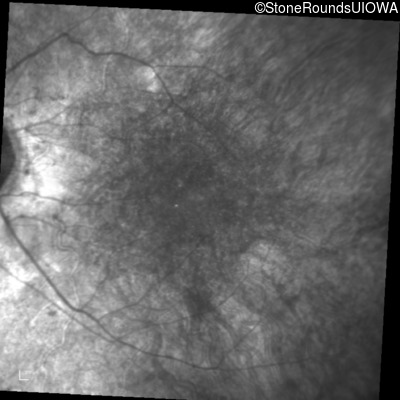
Infrared Fundus Photograph - Right - 20/60 +1 sc
Exemplar
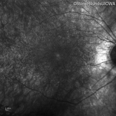
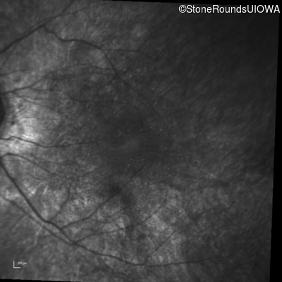
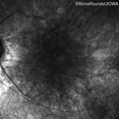
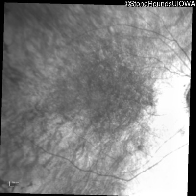
Infrared Fundus Photograph - Left - 20/50 -1 sc
Exemplar
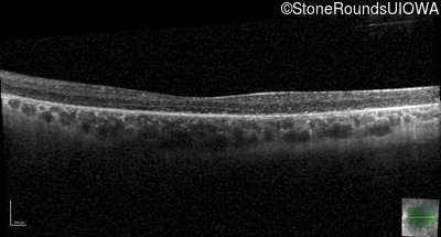
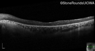
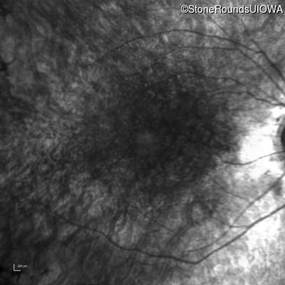
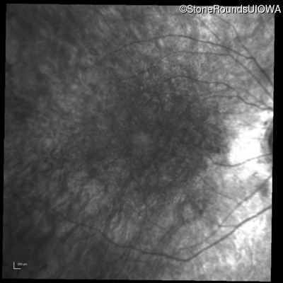
Visit at age: 31 years
Goldmann Visual Field - Right - 20/50 +1
Exemplar
Goldmann Visual Field - Left - 20/40 +1
Exemplar
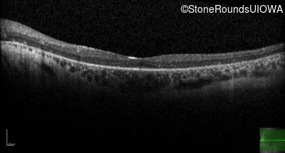
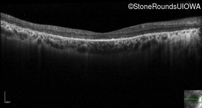
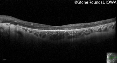
Optical Coherence Tomography - Right - 20/50 +1
Exemplar / OCT Stack
OCT Stack
OCT Stack
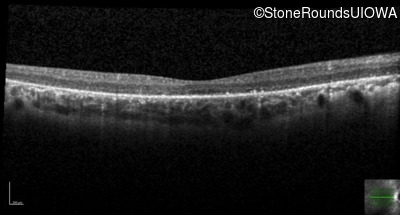
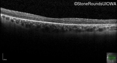
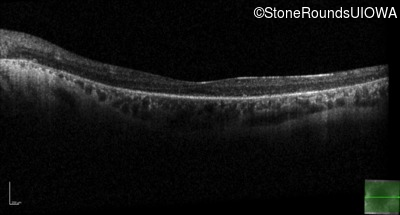
Optical Coherence Tomography - Left - 20/40 +1
Exemplar / OCT Stack
OCT Stack
OCT Stack
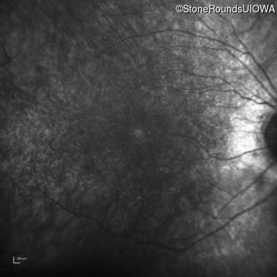
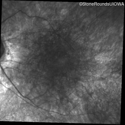
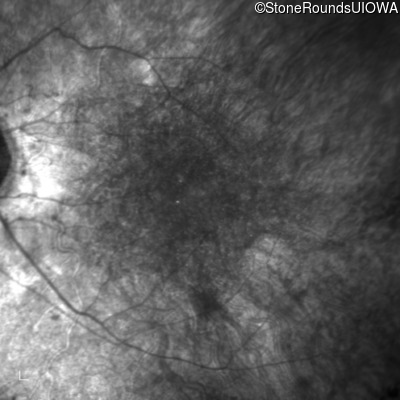
Infrared Fundus Photograph - Right - 20/50 +1
Exemplar

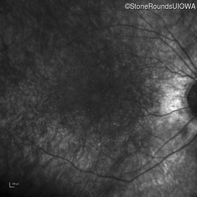
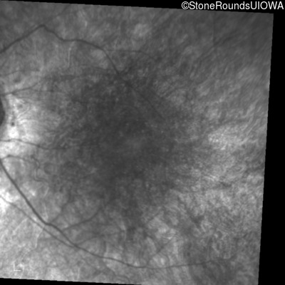
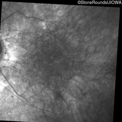
Infrared Fundus Photograph - Left - 20/40 +1
Exemplar

Visit at age: 33 years
Fundus Photography - Right - 20/50 -1
Exemplar
Fundus Photography - Left - 20/50 -2
Exemplar

Goldmann Visual Field - Right - 20/50 -1
Exemplar
Goldmann Visual Field - Left - 20/50 -2
Exemplar
Optical Coherence Tomography - Right - 20/50 -1
Exemplar / OCT Stack
OCT Stack
OCT Stack
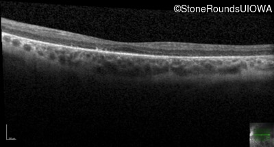
Optical Coherence Tomography - Left - 20/50 -2
Exemplar / OCT Stack
OCT Stack
OCT Stack
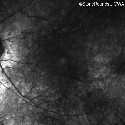
Infrared Fundus Photograph - Right - 20/50 -1
Exemplar
Infrared Fundus Photograph - Left - 20/50 -2
Exemplar
Visit at age: 36 years
Goldmann Visual Field - Right - 20/50 +1 sc
Exemplar
Goldmann Visual Field - Left - 20/50 +2 sc
Exemplar
Optical Coherence Tomography - Right - 20/50 +1 sc
Exemplar / OCT Stack
OCT Stack
OCT Stack
Optical Coherence Tomography - Left - 20/50 +2 sc
Exemplar / OCT Stack

OCT Stack
OCT Stack
Infrared Fundus Photograph - Right - 20/50 +1 sc
Exemplar
Infrared Fundus Photograph - Left - 20/50 +2 sc
Exemplar
Case Level Images
