Case
SR598
Student Mode
Stickler Syndrome (IIIE1)
Female
Female
Hidden
SR598
Student Mode
Stickler Syndrome (IIIE1)
Female
Female
Please Login or Register to download images or create a PowerPoint slideshow.
Visit at age: 28 years
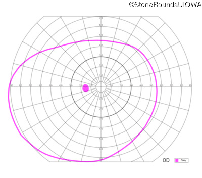
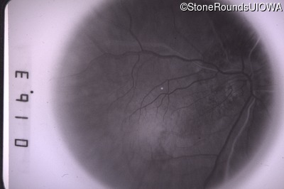
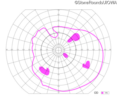
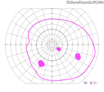
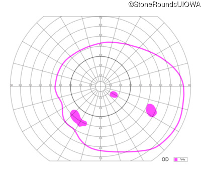
Goldmann Visual Field - Right - 20/50
Exemplar
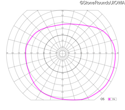
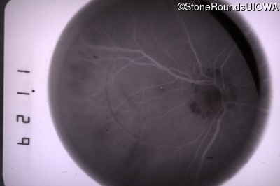
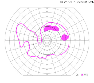
Goldmann Visual Field - Left - 20/40
Exemplar
Visit at age: 28 years (Visit 2)
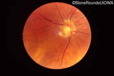
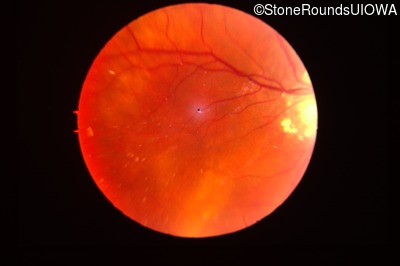
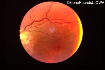
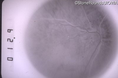
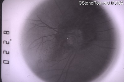
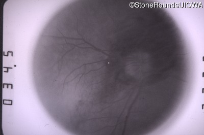
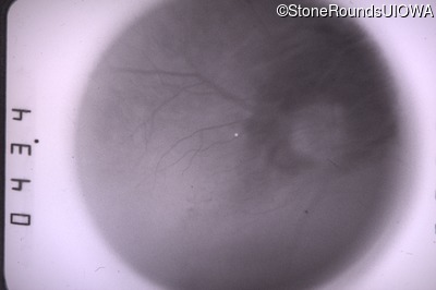
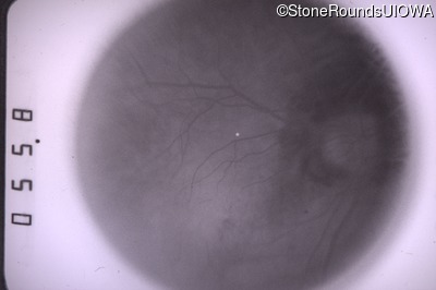
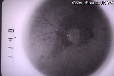
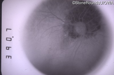
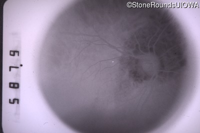
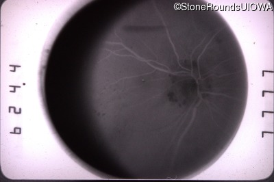
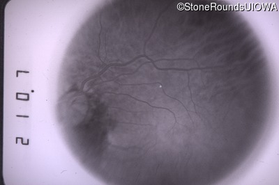
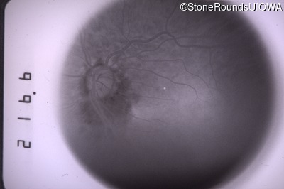
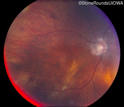
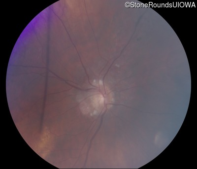
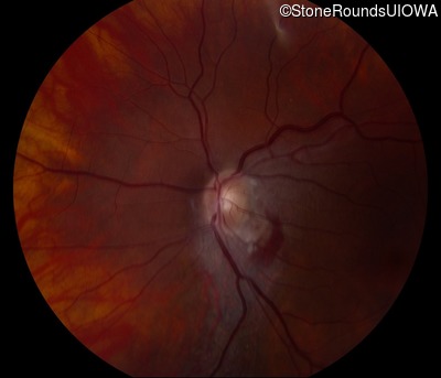
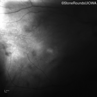
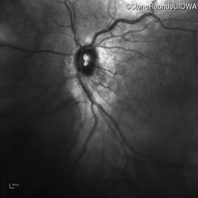
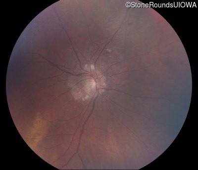
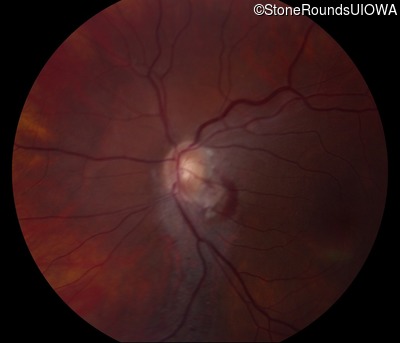
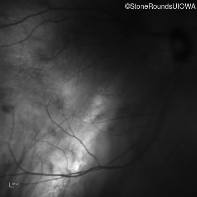
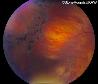
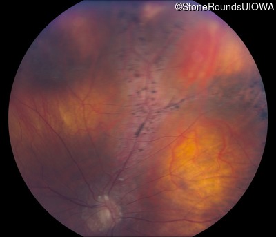
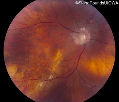
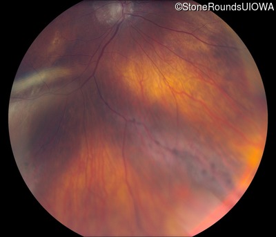
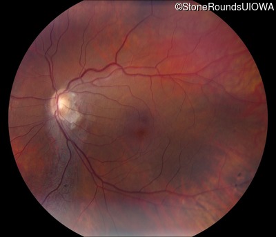
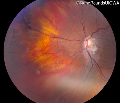
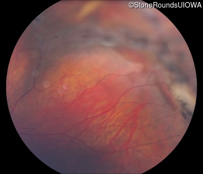
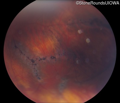
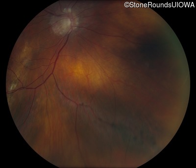
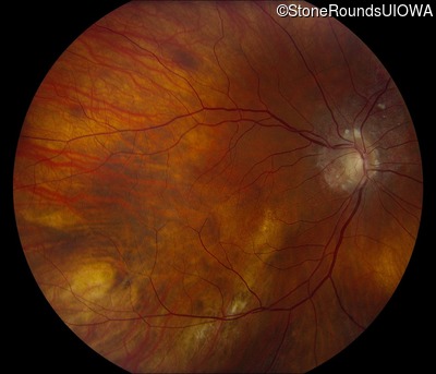
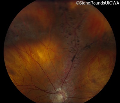
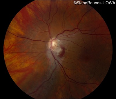
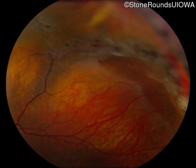
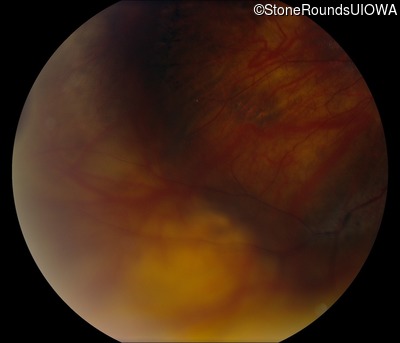
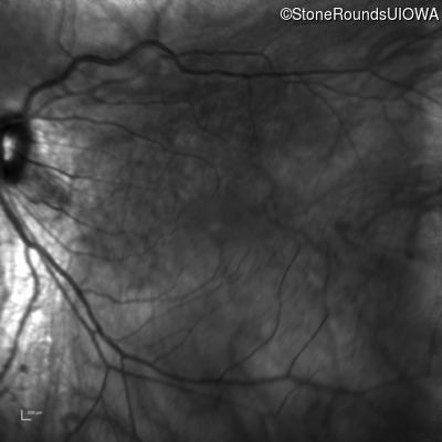
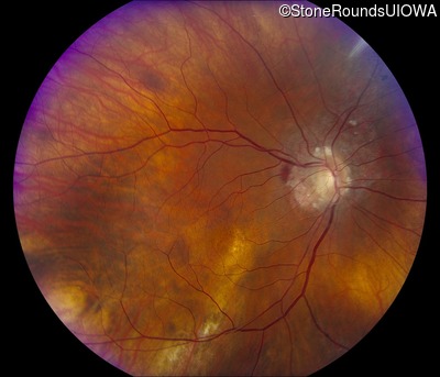
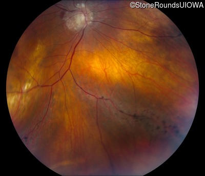
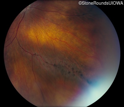
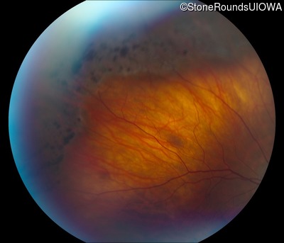
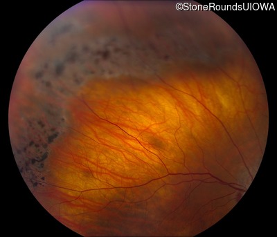
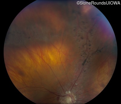
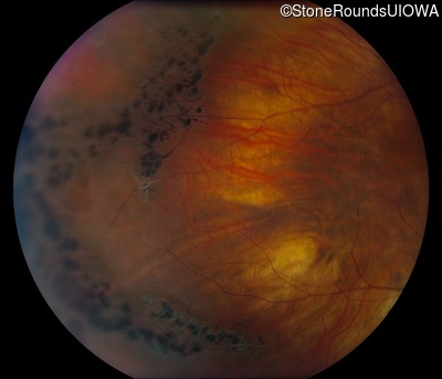
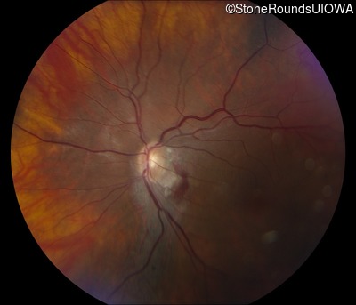
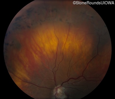
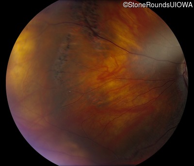
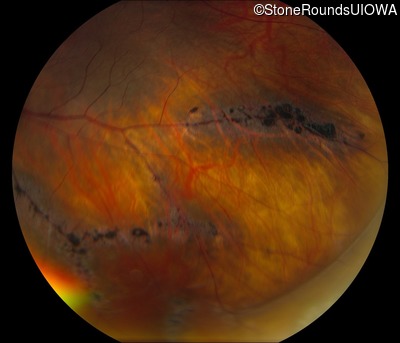
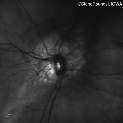
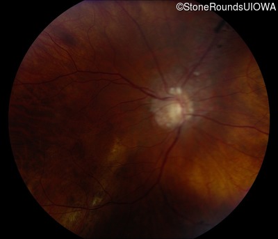
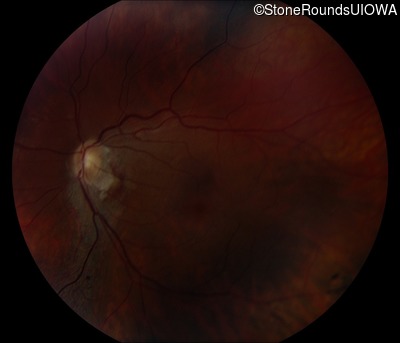
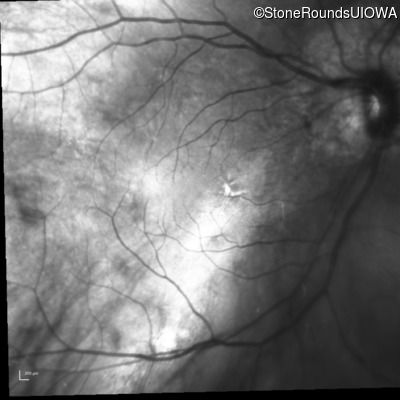
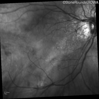
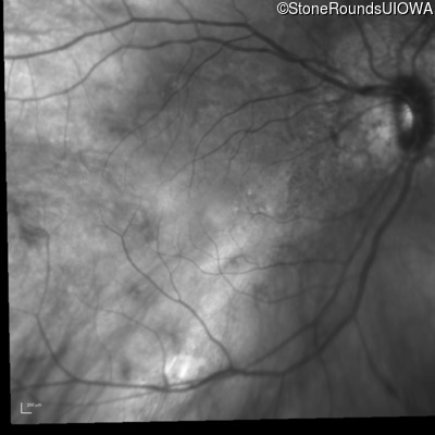
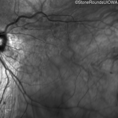
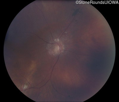
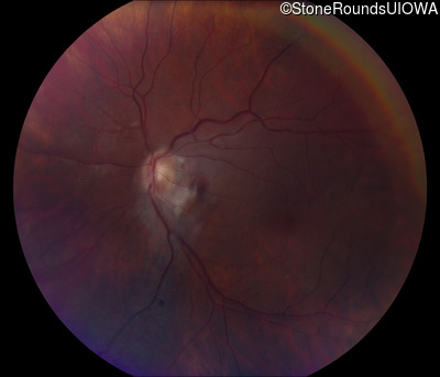
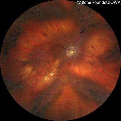
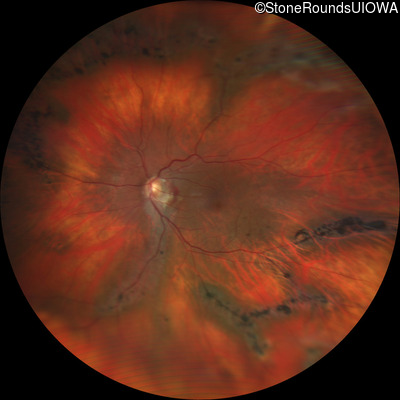
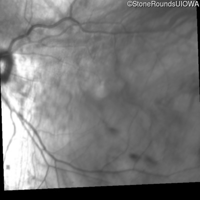
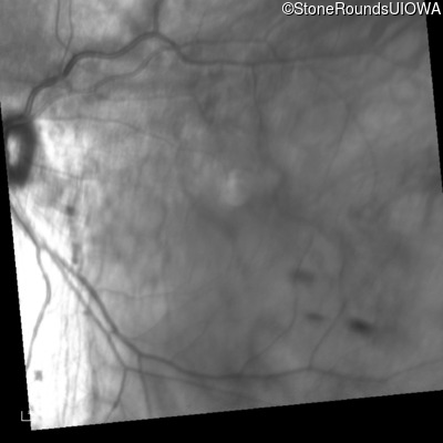
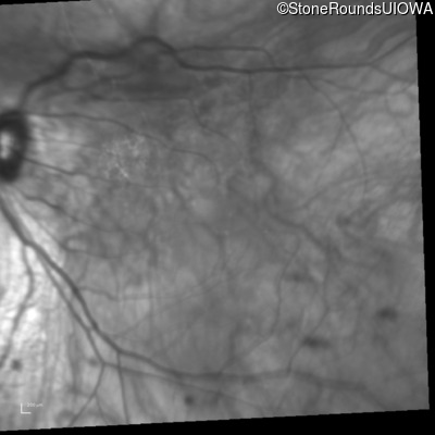
Fundus Photography - Right - 20/60 -2
Exemplar
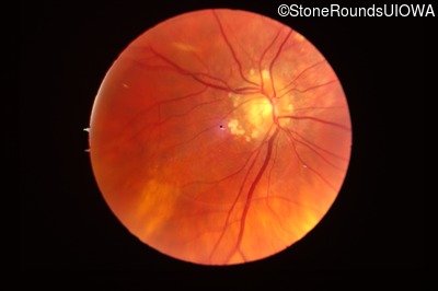
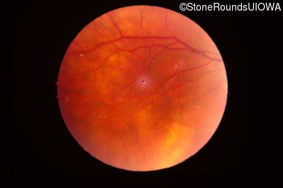
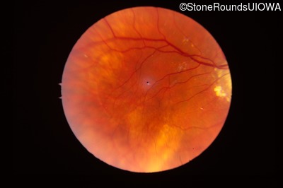
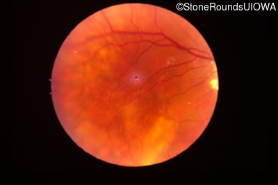
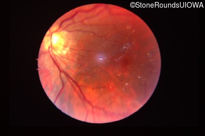
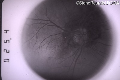
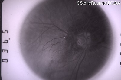
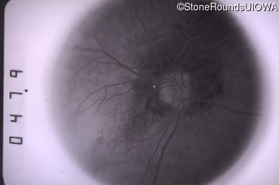
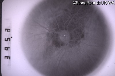
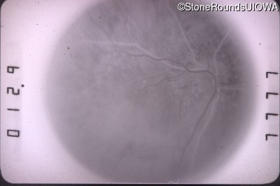
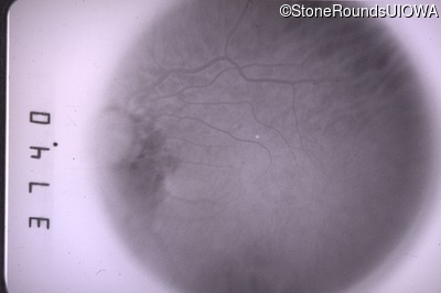
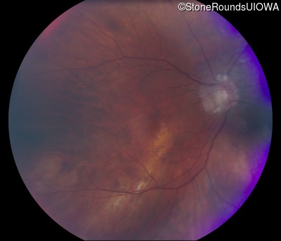
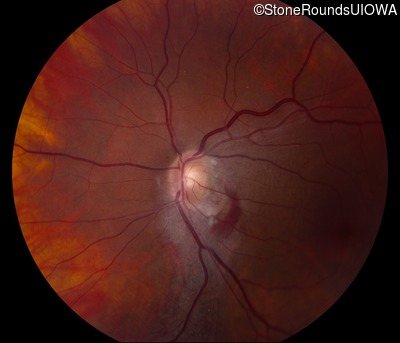
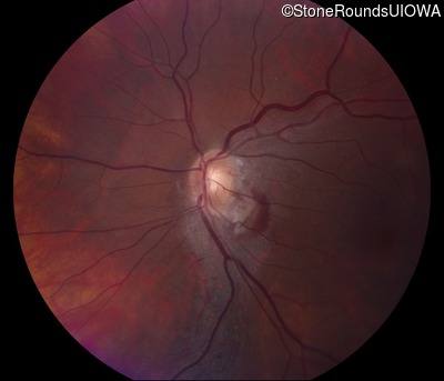
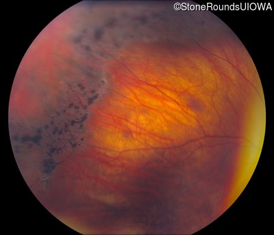
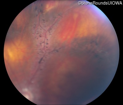
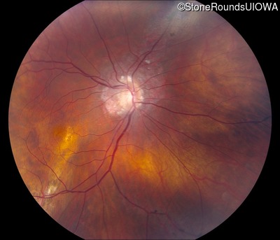
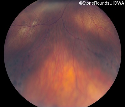
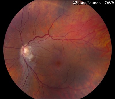
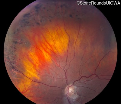
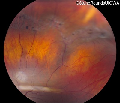
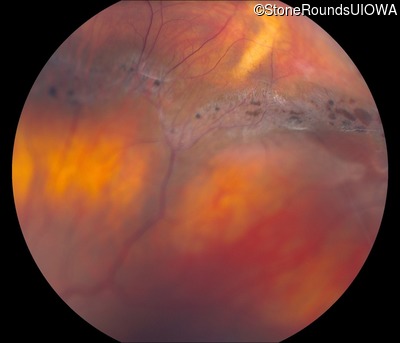
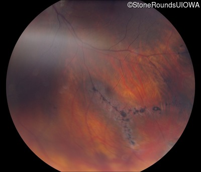
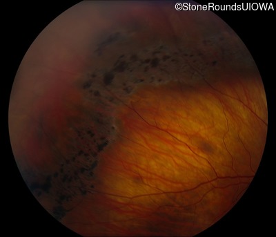
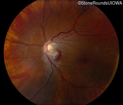
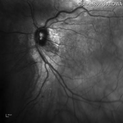
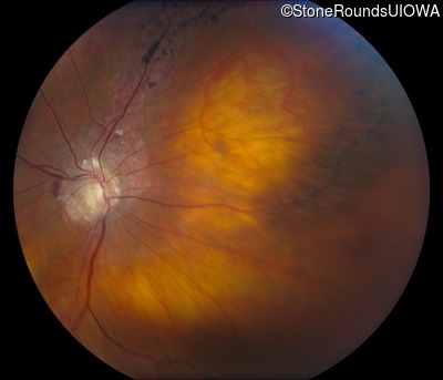
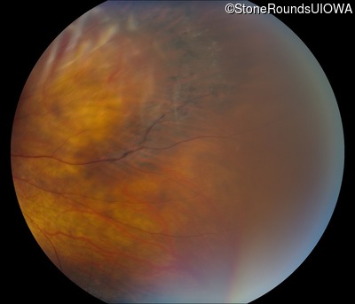
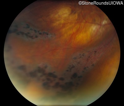
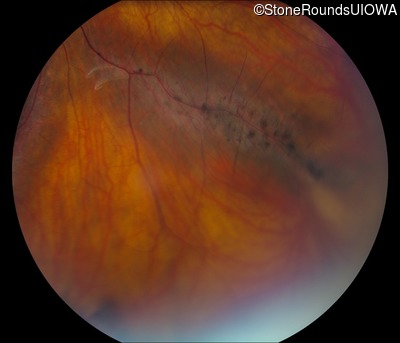
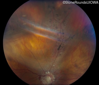
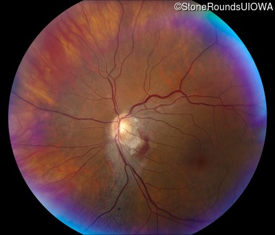
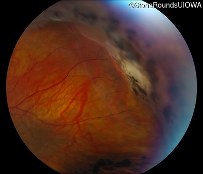
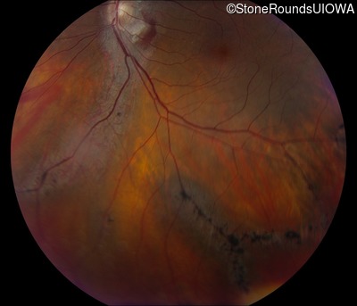
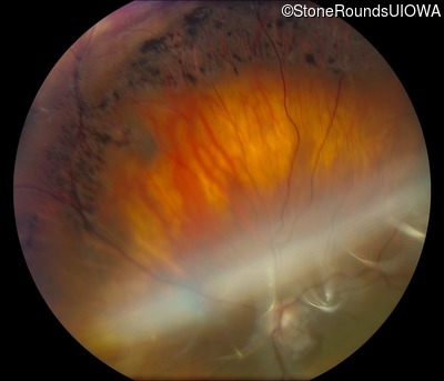
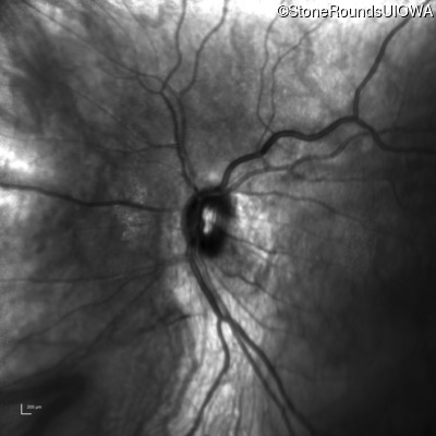
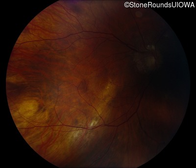
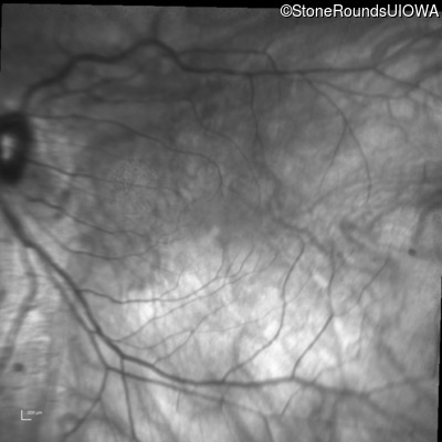
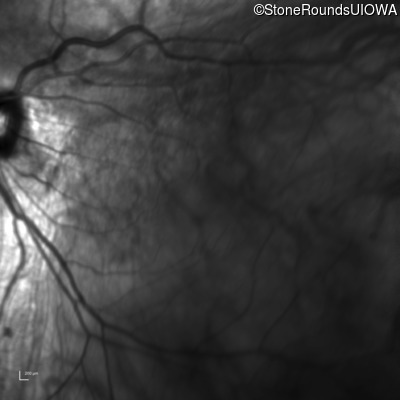
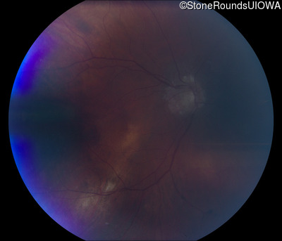
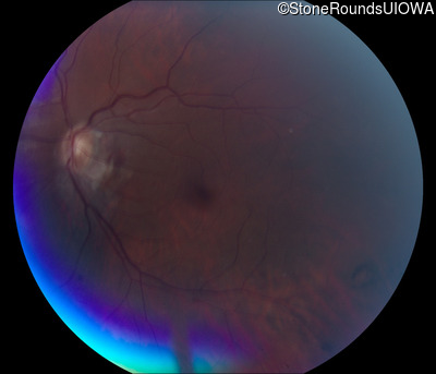
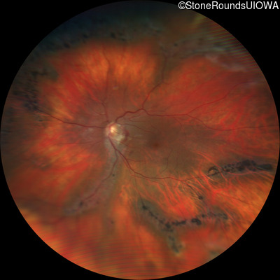
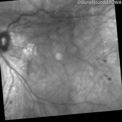
Fundus Photography - Left - 20/30
Exemplar
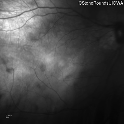
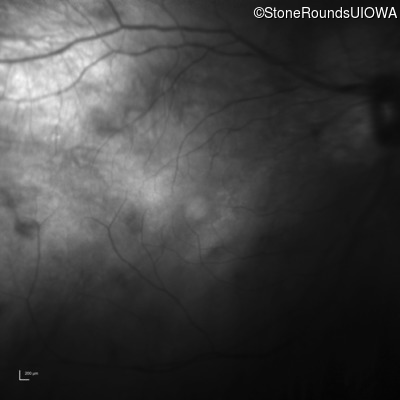
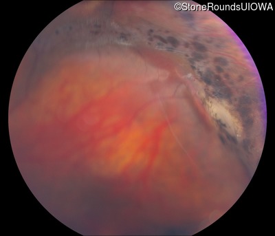
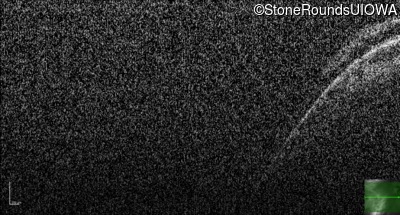
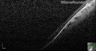
Fluorescein Angiography - Right - 20/60 -2
Exemplar

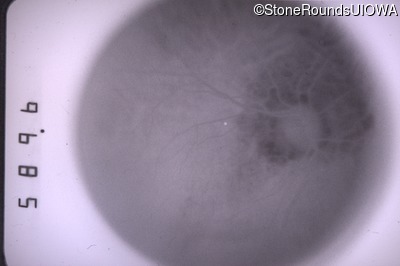
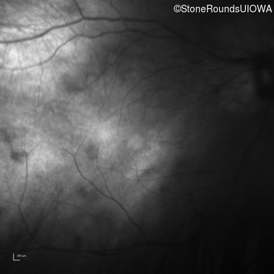
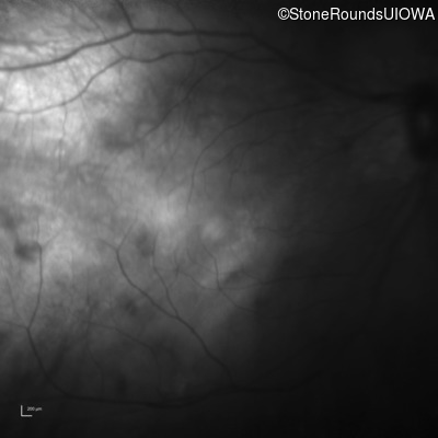
Fluorescein Angiography - Left - 20/30
Exemplar
Visit at age: 29 years
Goldmann Visual Field - Right - 20/40
Exemplar
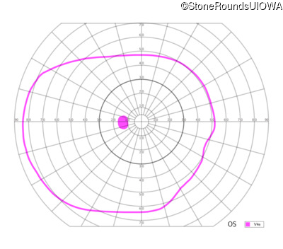
Goldmann Visual Field - Left - 20/50
Exemplar
Visit at age: 32 years
Goldmann Visual Field - Right - 20/30 -2
Exemplar
Goldmann Visual Field - Left - 20/25 -2
Exemplar
Visit at age: 36 years
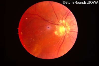
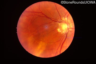
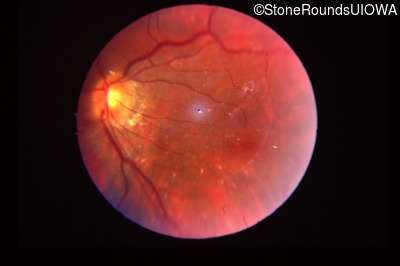
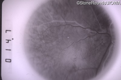
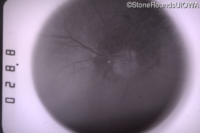
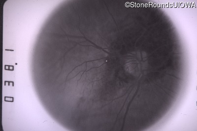
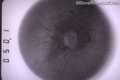
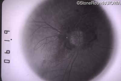
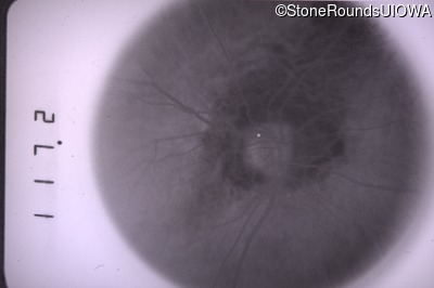
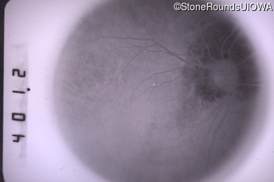
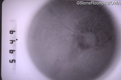
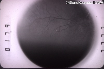
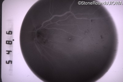
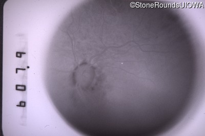
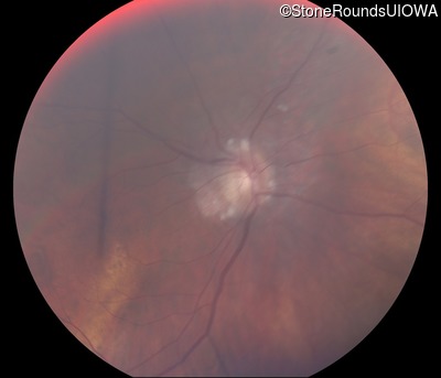
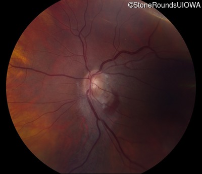
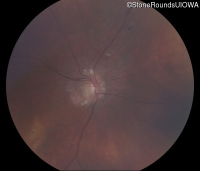
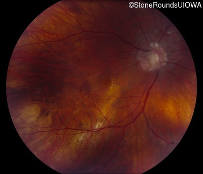
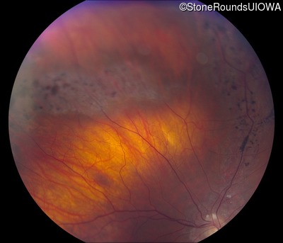
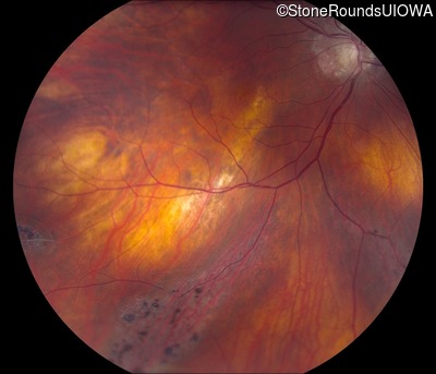
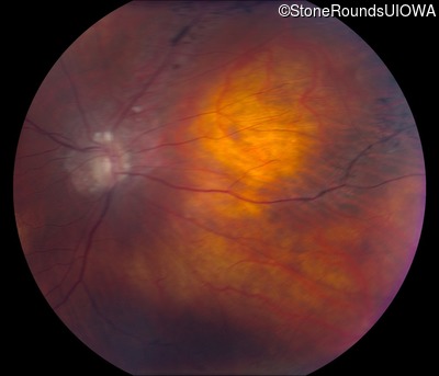
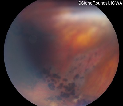
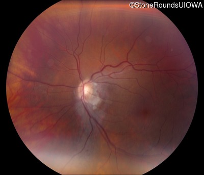
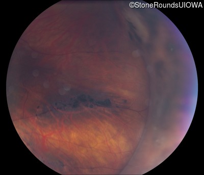
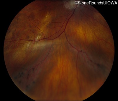
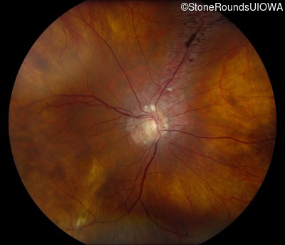
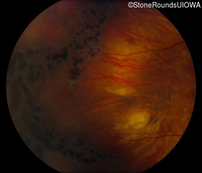
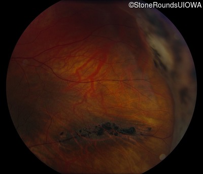
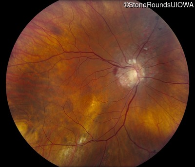
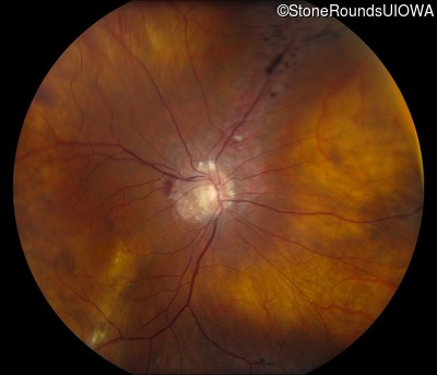
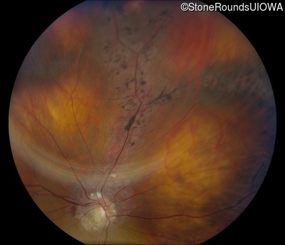
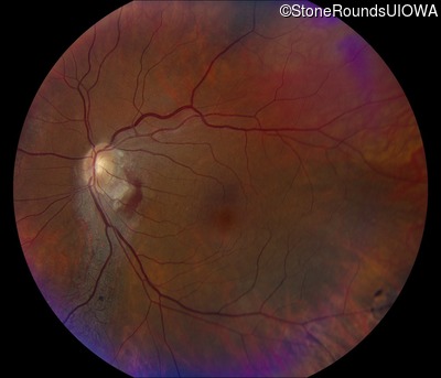
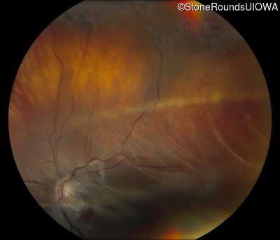
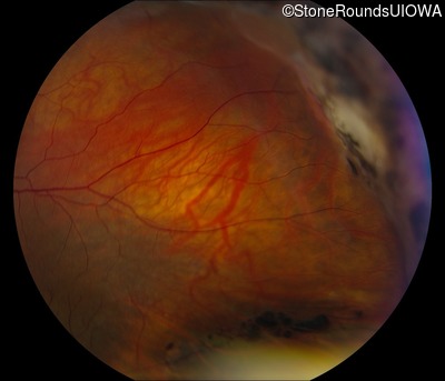
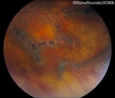
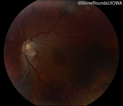
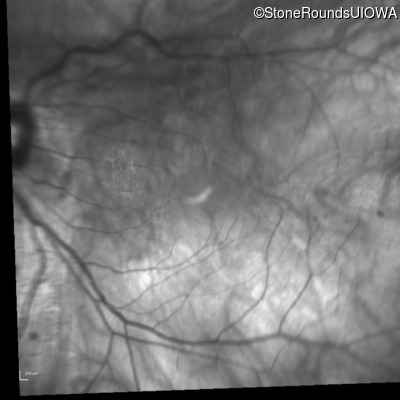
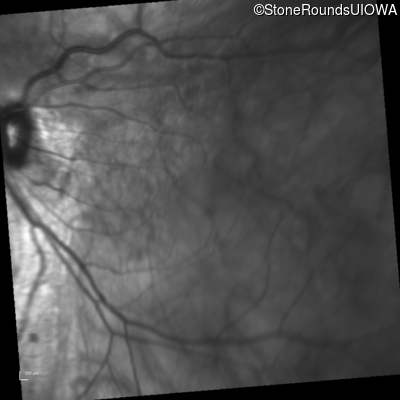
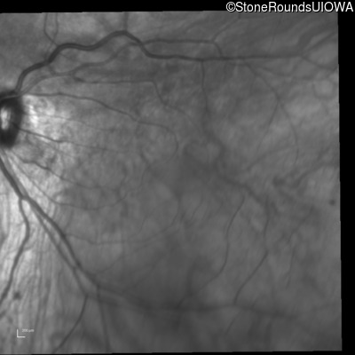
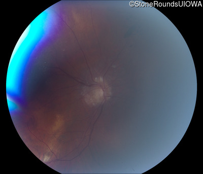
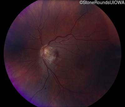
Fundus Photography - Right - 20/40 +2
Exemplar
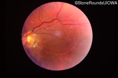
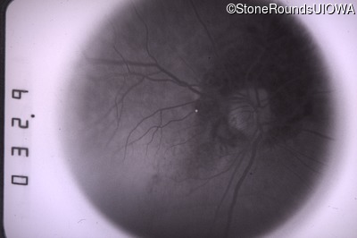
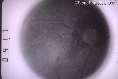
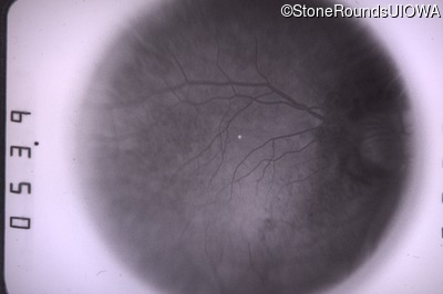
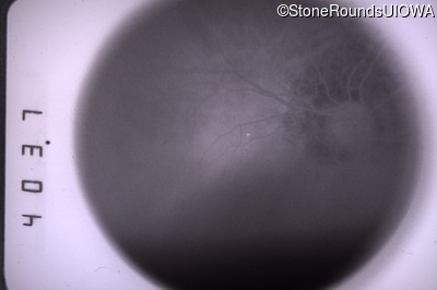
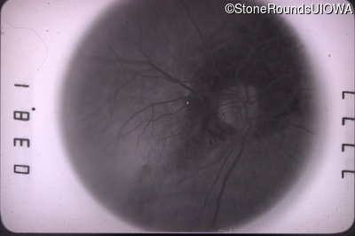
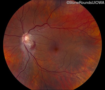
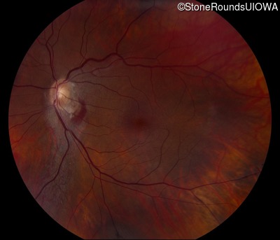
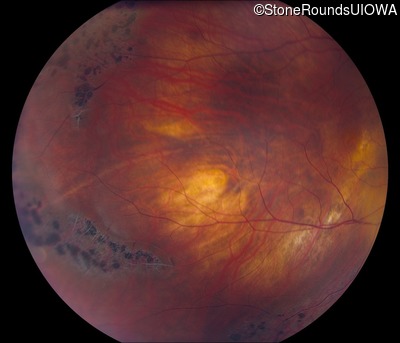
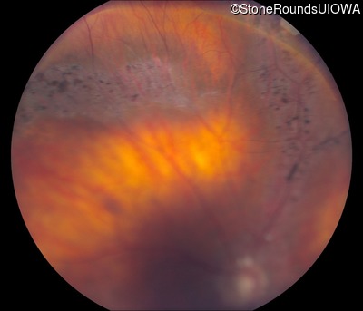
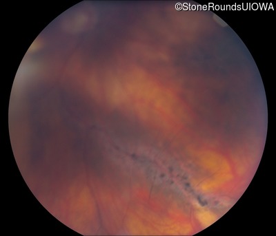
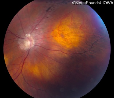
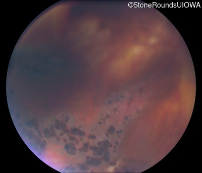
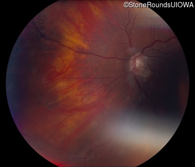
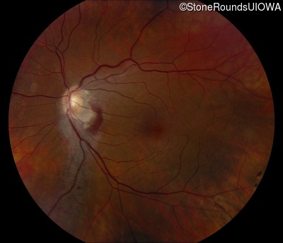
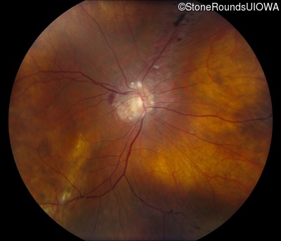
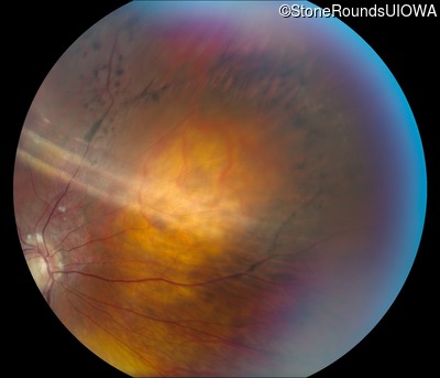
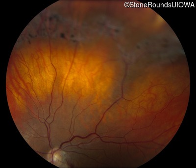
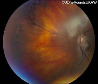
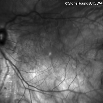
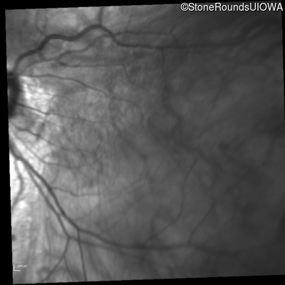
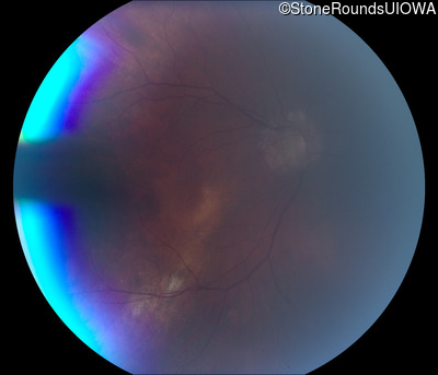
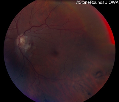
Fundus Photography - Left - 20/25 +2
Exemplar
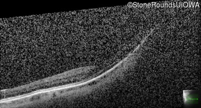
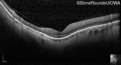
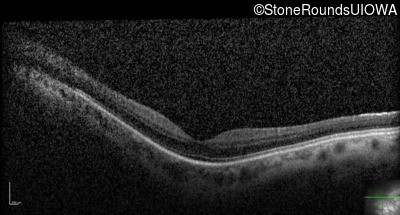
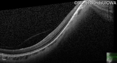
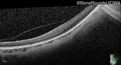
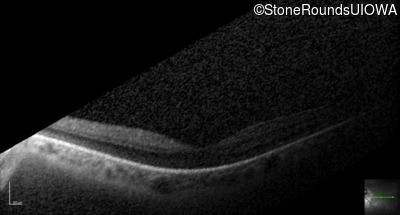
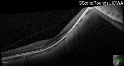
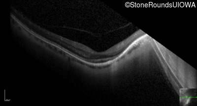
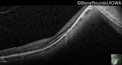
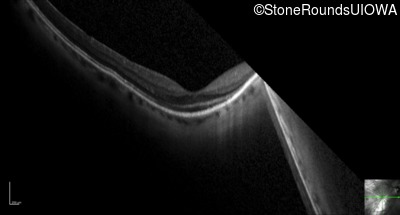
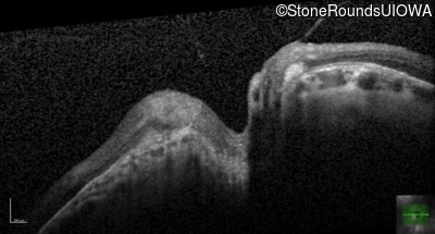
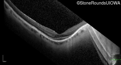
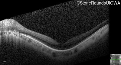
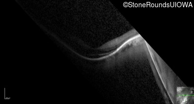
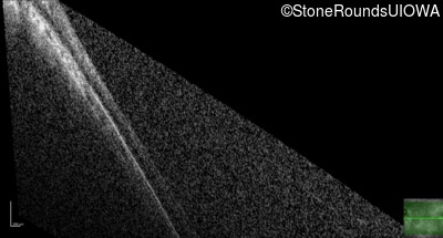
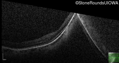
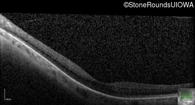
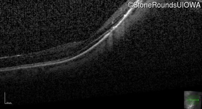
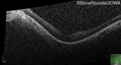
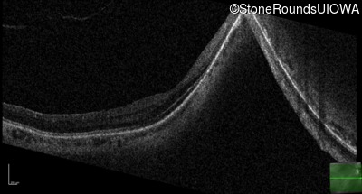
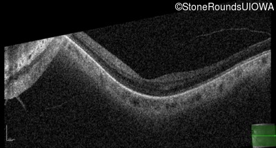
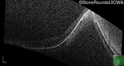
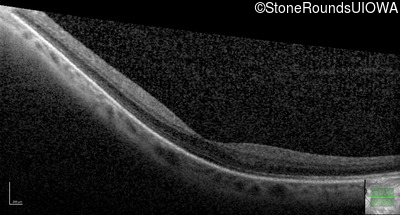
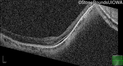
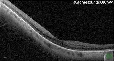
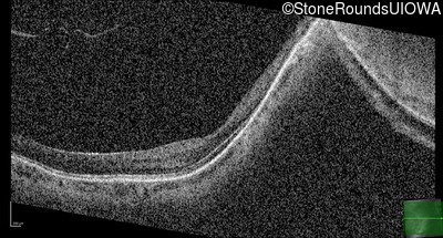
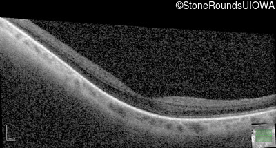
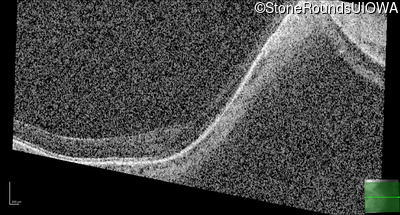
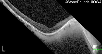
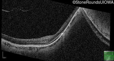
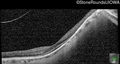
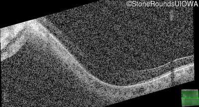
Optical Coherence Tomography - Right - 20/40 +2
Exemplar / OCT Stack
OCT Stack
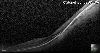
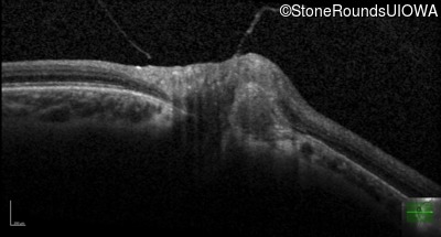
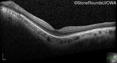
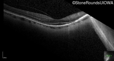
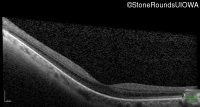
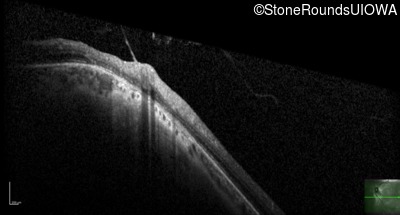
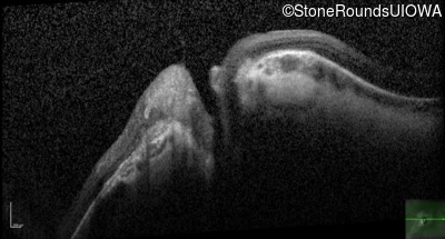
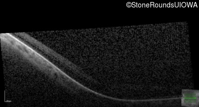
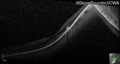
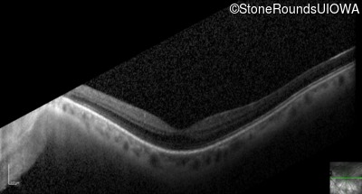
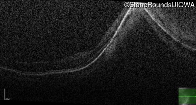
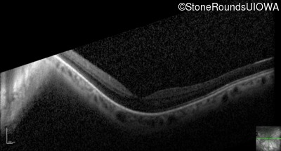
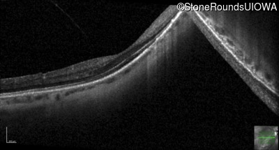
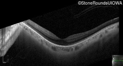
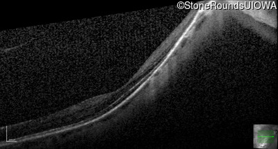
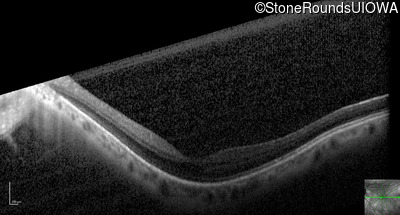
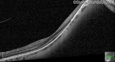
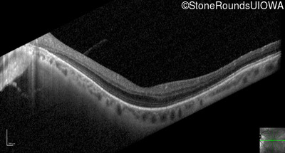
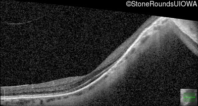
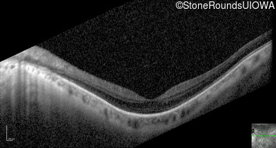
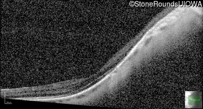
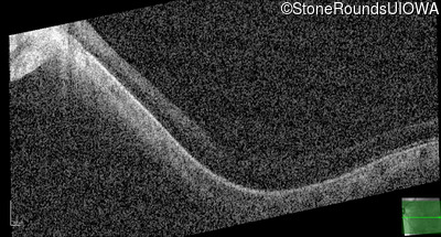
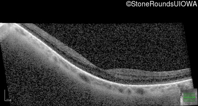
Optical Coherence Tomography - Left - 20/25 +2
Exemplar / OCT Stack
OCT Stack
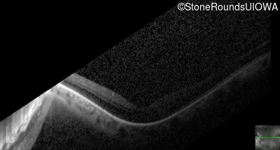
OCT Stack
OCT Stack
OCT Stack
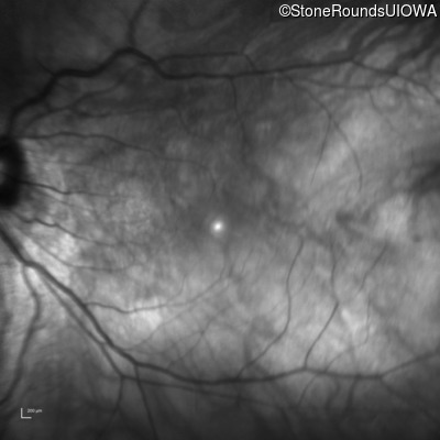
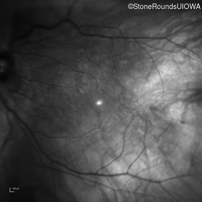
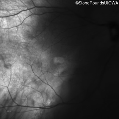
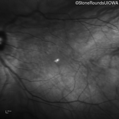
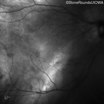
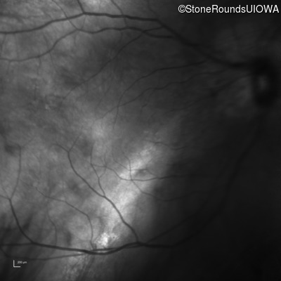
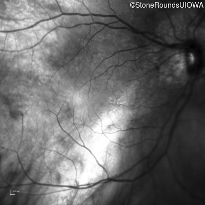
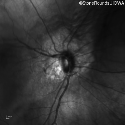
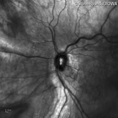
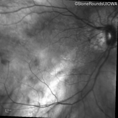
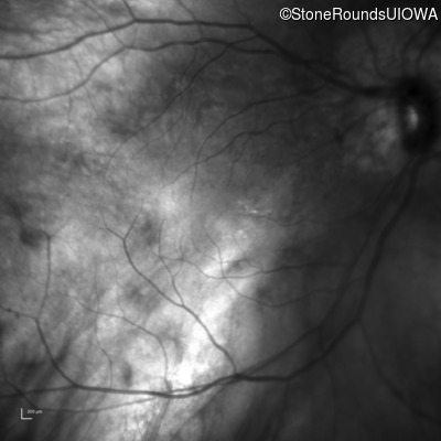
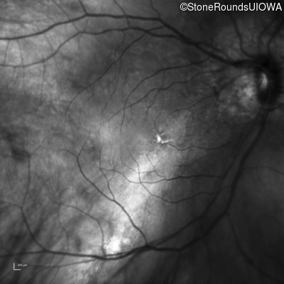
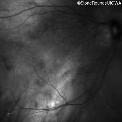
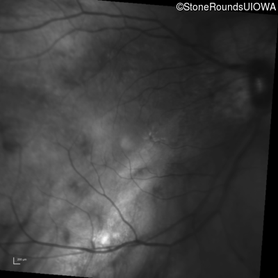
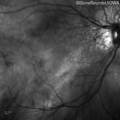
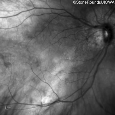
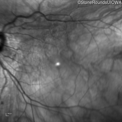
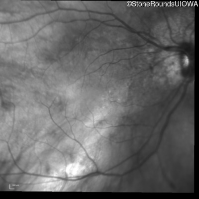
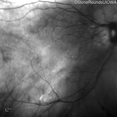
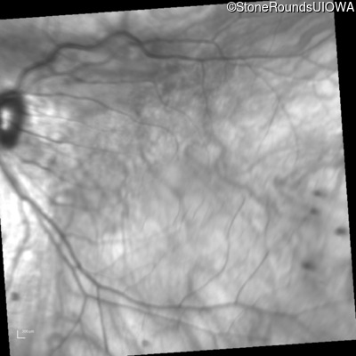
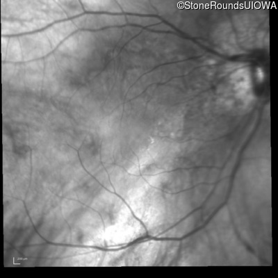
Infrared Fundus Photograph - Right - 20/40 +2
Exemplar
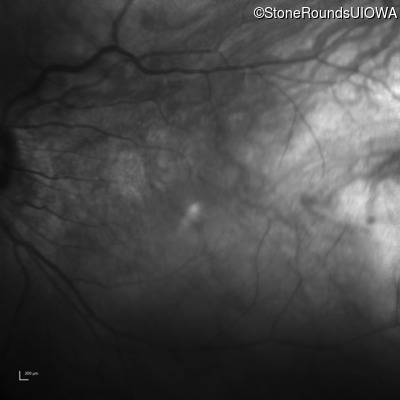
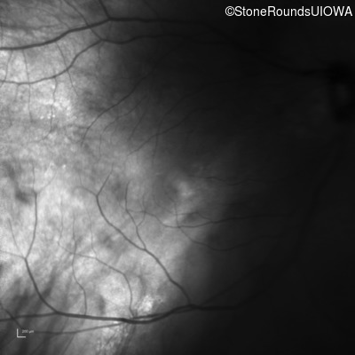
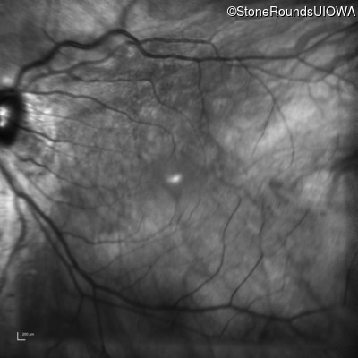
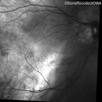
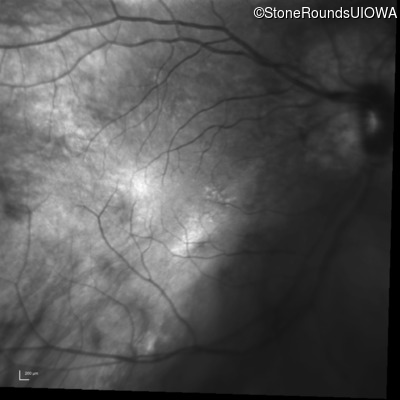
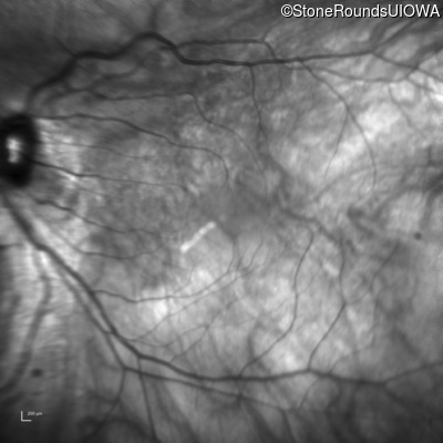
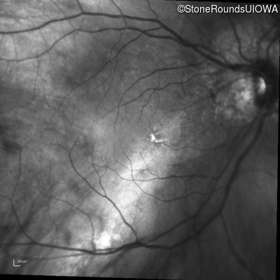
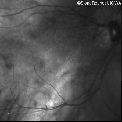
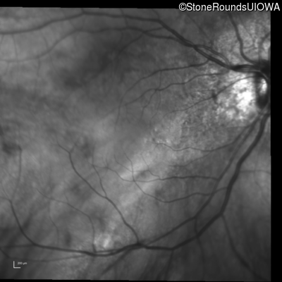
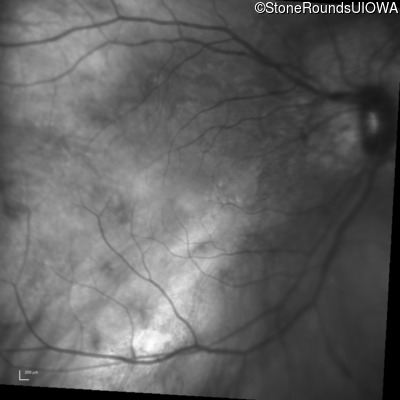
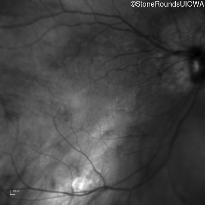
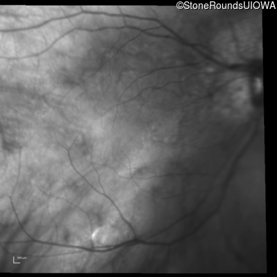
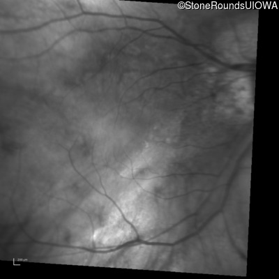
Infrared Fundus Photograph - Left - 20/25 +2
Exemplar
Visit at age: 37 years
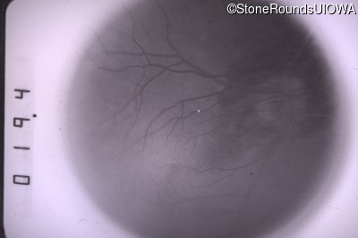
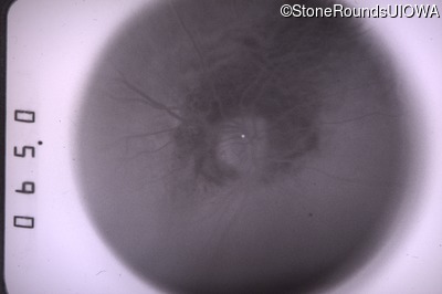
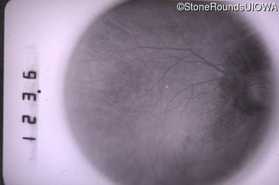
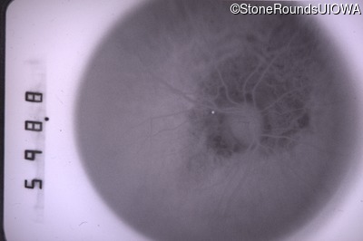
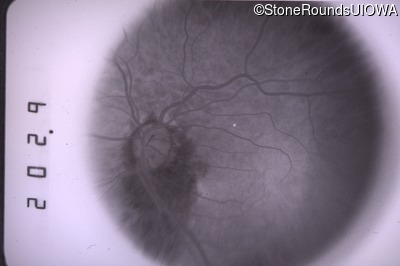
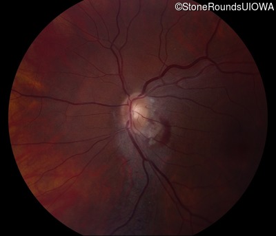
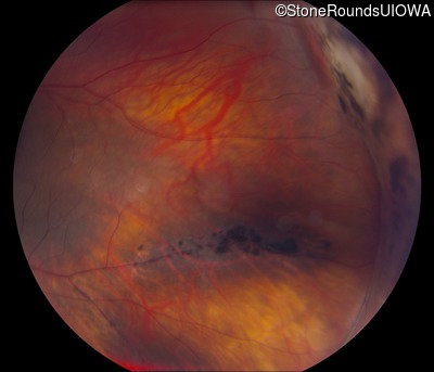
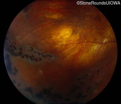
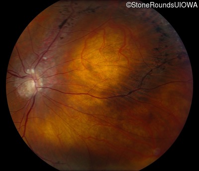
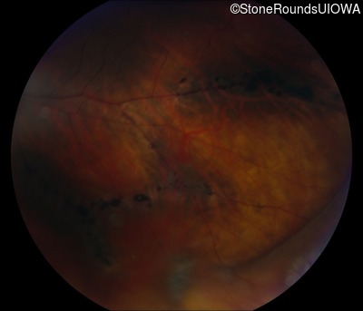
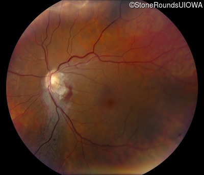
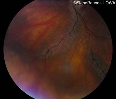
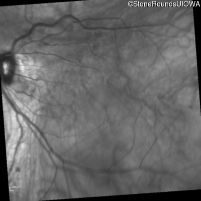
Fundus Photography - Right - 20/30
Exemplar
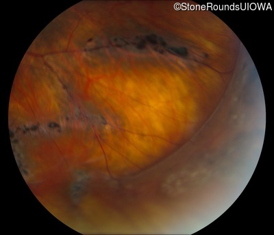
Fundus Photography - Left - 20/25 -2
Exemplar
Goldmann Visual Field - Right - 20/30
Exemplar
Goldmann Visual Field - Left - 20/25 -2
Exemplar

Visit at age: 38 years
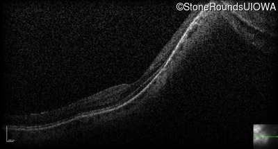
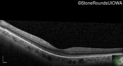
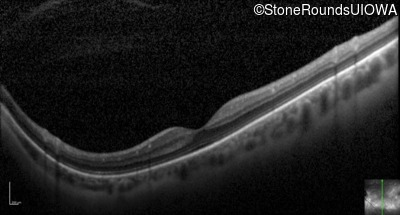
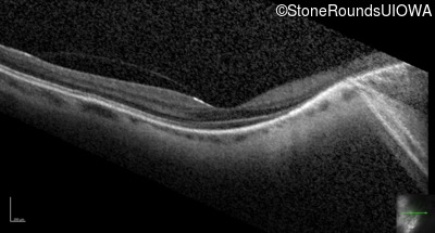
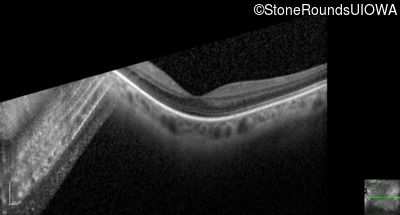
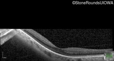
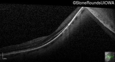
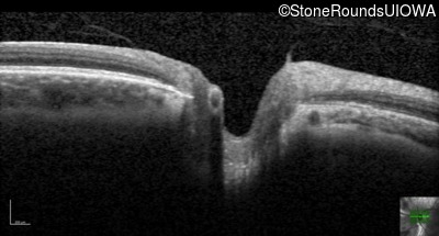
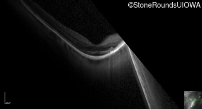
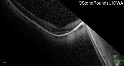
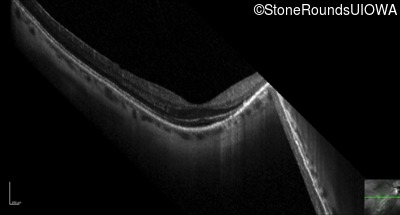
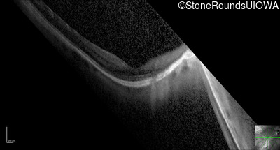
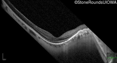
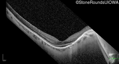
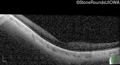
Optical Coherence Tomography - Right - 20/25 -3
Exemplar / OCT Stack
OCT Stack
OCT Stack
OCT Stack
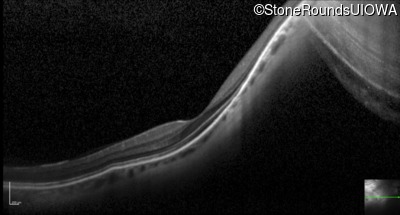
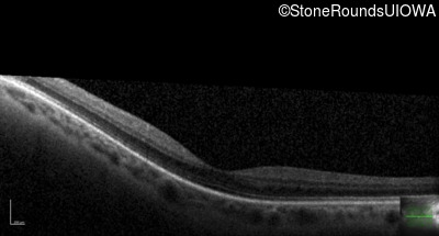
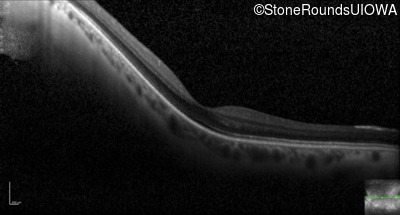
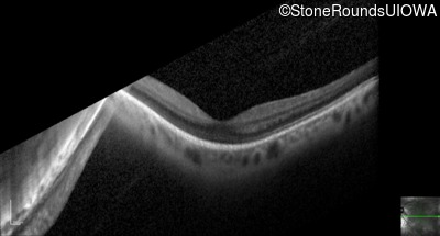
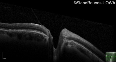
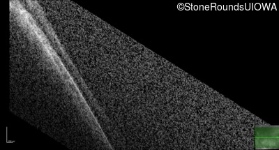
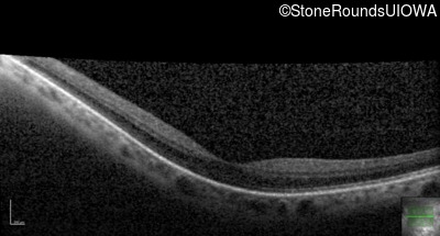
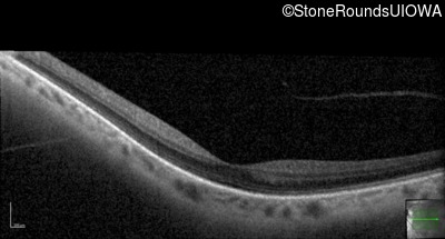
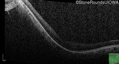
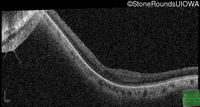
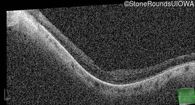
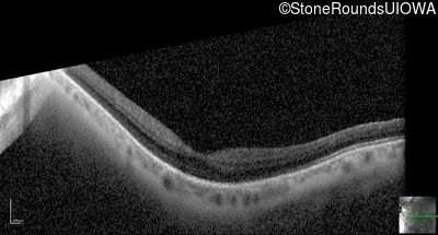
Optical Coherence Tomography - Left - 20/25 -2
Exemplar / OCT Stack
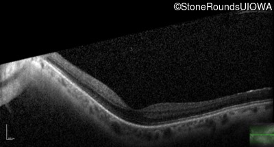
OCT Stack
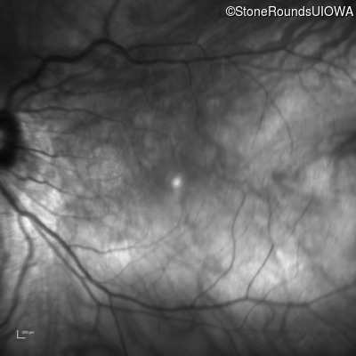
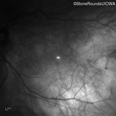
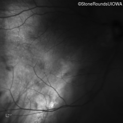
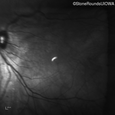
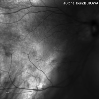
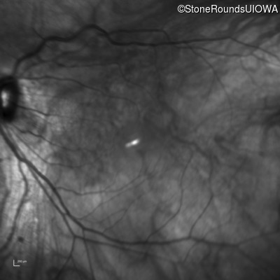
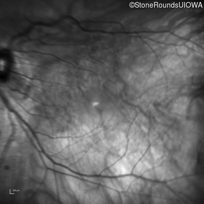
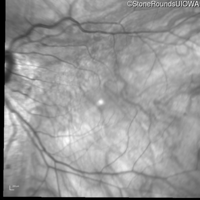
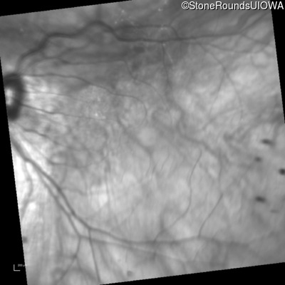
Infrared Fundus Photograph - Right - 20/25 -3
Exemplar
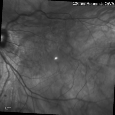
Infrared Fundus Photograph - Left - 20/25 -2
Exemplar
Visit at age: 39 years
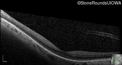
Optical Coherence Tomography - Right - 20/30 +2
Exemplar / OCT Stack
OCT Stack
Optical Coherence Tomography - Left - 20/30 +1
Exemplar / OCT Stack
OCT Stack
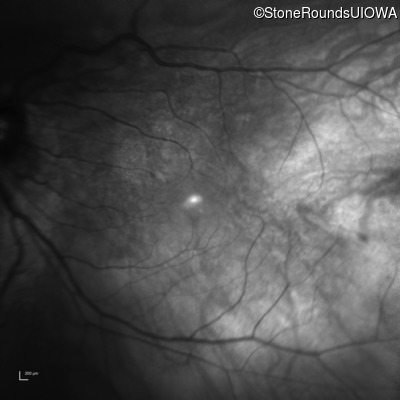
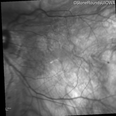
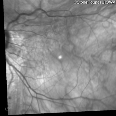
Infrared Fundus Photograph - Right - 20/30 +2
Exemplar
Infrared Fundus Photograph - Left - 20/30 +1
Exemplar

Visit at age: 41 years
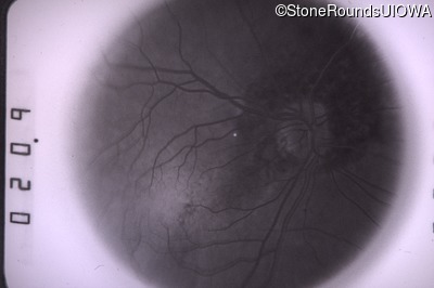
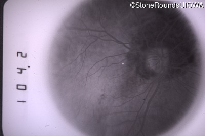
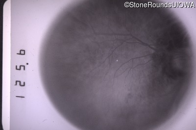
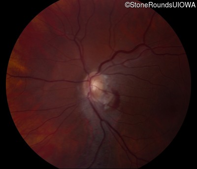
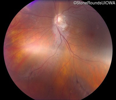
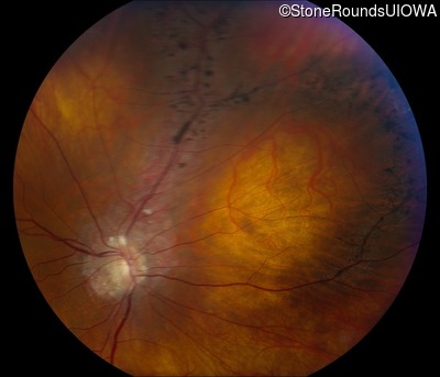
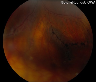
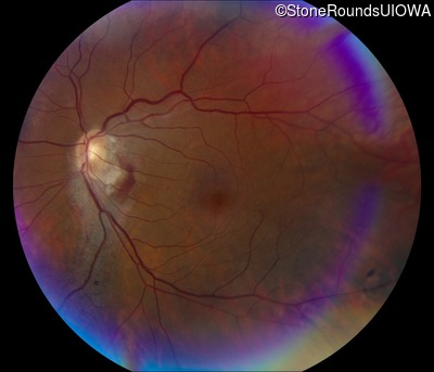
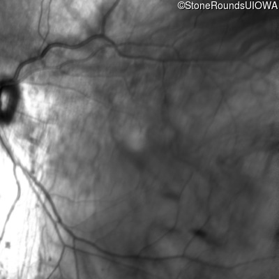
Fundus Photography - Right - 20/30 +2
Exemplar
Fundus Photography - Left - 20/30 +2
Exemplar
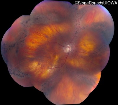
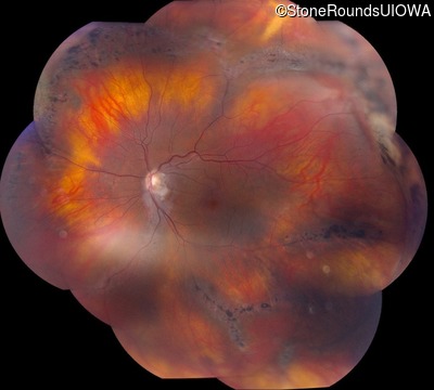
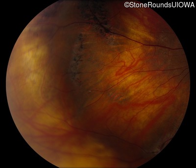
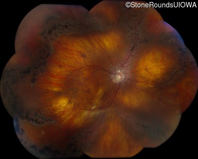
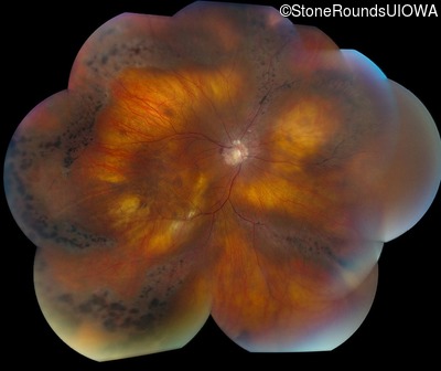
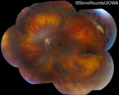
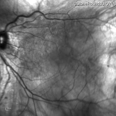
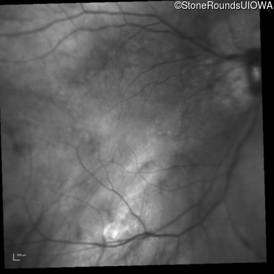
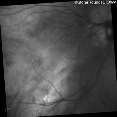
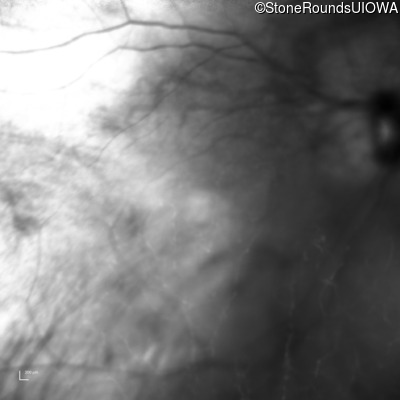
Fundus Montage - Right - 20/30 +2
Exemplar
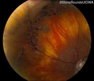
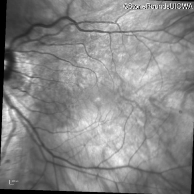
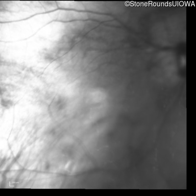
Fundus Montage - Left - 20/30 +2
Exemplar
Visit at age: 41 years (Visit 2)
Fundus Photography - Right - 20/32 -1
Exemplar


Fundus Photography - Left - 20/32
Exemplar


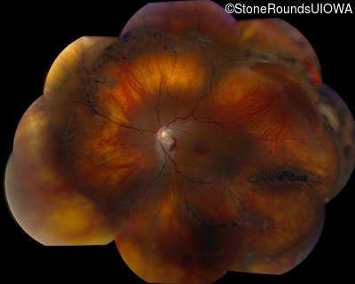
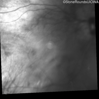
Fundus Montage - Right - 20/32 -1
Exemplar
Fundus Montage - Left - 20/32
Exemplar
Optical Coherence Tomography - Right - 20/32 -1
Exemplar / OCT Stack
OCT Stack
Optical Coherence Tomography - Left - 20/32
Exemplar / OCT Stack
OCT Stack
OCT Stack
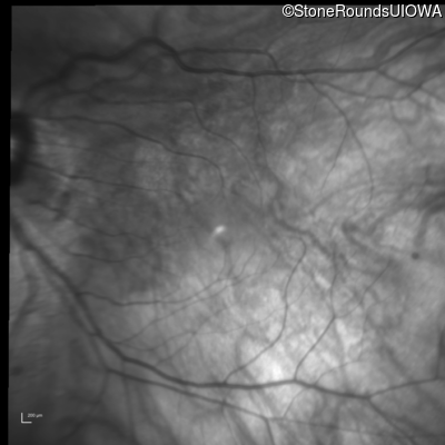
Infrared Fundus Photograph - Right - 20/32 -1
Exemplar
Infrared Fundus Photograph - Left - 20/32
Exemplar

Visit at age: 42 years
Fundus Photography - Right - 20/40 +2
Exemplar
Fundus Photography - Left - 20/30 +2
Exemplar
Fundus Montage - Right - 20/40 +2
Exemplar
Fundus Montage - Left - 20/30 +2
Exemplar
Optical Coherence Tomography - Right - 20/40 +2
Exemplar / OCT Stack
OCT Stack
OCT Stack
OCT Stack
Optical Coherence Tomography - Left - 20/30 +2
Exemplar / OCT Stack
OCT Stack
OCT Stack
OCT Stack
Infrared Fundus Photograph - Right - 20/40 +2
Exemplar
Infrared Fundus Photograph - Left - 20/30 +2
Exemplar
Visit at age: 42 years (Visit 2)
Fundus Photography - Right - 20/30 -2
Exemplar
Fundus Photography - Left - 20/25 -2
Exemplar
Optical Coherence Tomography - Right - 20/30 -2
Exemplar / OCT Stack
OCT Stack
OCT Stack
Optical Coherence Tomography - Left - 20/25 -2
Exemplar / OCT Stack
OCT Stack
OCT Stack
Infrared Fundus Photograph - Right - 20/30 -2
Exemplar

Infrared Fundus Photograph - Left - 20/25 -2
Exemplar
Visit at age: 43 years
Optical Coherence Tomography - Right - 20/25 -1
Exemplar / OCT Stack
OCT Stack
OCT Stack
Optical Coherence Tomography - Left - 20/30 -2
Exemplar / OCT Stack
OCT Stack
OCT Stack
Infrared Fundus Photograph - Right - 20/25 -1
Exemplar
Infrared Fundus Photograph - Left - 20/30 -2
Exemplar
Visit at age: 44 years
Optical Coherence Tomography - Right - 20/30 -1
Exemplar / OCT Stack
OCT Stack
OCT Stack
Optical Coherence Tomography - Left - 20/40
Exemplar / OCT Stack
OCT Stack
OCT Stack
Infrared Fundus Photograph - Right - 20/30 -1
Exemplar
Infrared Fundus Photograph - Left - 20/40
Exemplar
Visit at age: 45 years
Optical Coherence Tomography - Right - 20/20 -3
Exemplar / OCT Stack
OCT Stack
OCT Stack
Optical Coherence Tomography - Left - 20/25 +2
Exemplar / OCT Stack
OCT Stack
OCT Stack
Infrared Fundus Photograph - Right - 20/20 -3
Exemplar
Infrared Fundus Photograph - Left - 20/25 +2
Exemplar
Visit at age: 46 years
Optical Coherence Tomography - Right - 20/25 -3
Exemplar / OCT Stack
OCT Stack
OCT Stack
Optical Coherence Tomography - Left - 20/30 +1
Exemplar / OCT Stack
OCT Stack
OCT Stack
Infrared Fundus Photograph - Right - 20/25 -3
Exemplar
Infrared Fundus Photograph - Left - 20/30 +1
Exemplar
Visit at age: 47 years
Optical Coherence Tomography - Right - 20/30 +1
Exemplar / OCT Stack
OCT Stack
OCT Stack
Optical Coherence Tomography - Left - 20/25 +2
Exemplar / OCT Stack
OCT Stack
OCT Stack
Infrared Fundus Photograph - Right - 20/30 +1
Exemplar
Infrared Fundus Photograph - Left - 20/25 +2
Exemplar
Visit at age: 47 years (Visit 2)
Fundus Photography - Right - 20/32 -1
Exemplar
Fundus Photography - Left - 20/32
Exemplar
Optical Coherence Tomography - Right - 20/32 -1
Exemplar / OCT Stack
OCT Stack
OCT Stack
Optical Coherence Tomography - Left - 20/32
Exemplar / OCT Stack
OCT Stack
OCT Stack
Infrared Fundus Photograph - Right - 20/32 -1
Exemplar
Infrared Fundus Photograph - Left - 20/32
Exemplar

Visit at age: 48 years
Fundus Photography - Right - 20/40 +1
Exemplar
Fundus Photography - Left - 20/40 +1
Exemplar
Optical Coherence Tomography - Right - 20/40 +1
Exemplar / OCT Stack
OCT Stack
OCT Stack
Optical Coherence Tomography - Left - 20/40 +1
Exemplar / OCT Stack
OCT Stack
OCT Stack
Infrared Fundus Photograph - Right - 20/40 +1
Exemplar
Infrared Fundus Photograph - Left - 20/40 +1
Exemplar
Visit at age: 49 years
Optical Coherence Tomography - Right - 20/40 +2
Exemplar / OCT Stack
OCT Stack
Optical Coherence Tomography - Left - 20/32 +1
Exemplar / OCT Stack
OCT Stack
Infrared Fundus Photograph - Right - 20/40 +2
Exemplar
Infrared Fundus Photograph - Left - 20/32 +1
Exemplar
Case Level Images
