Case
SR1066
Student Mode
AD Cone and Cone Rod Dystrophy (IA1bii)
Male
Male
Hidden
SR1066
Student Mode
AD Cone and Cone Rod Dystrophy (IA1bii)
Male
Male


History
This 30 year old man experienced gradually decreasing visual acuity and increasing photophobia beginning at around ten years of age. As a young child, his best corrected visual acuity was near normal (20/30 OU).
| Color Vision: | Could see the control pseudoisochromatic plate but none of the test plates with either eye. |
|---|---|
| Refraction OD: | -5.25 +1.25 x 120 |
| Refraction OS: | -4.25 +1.25 x 030 |
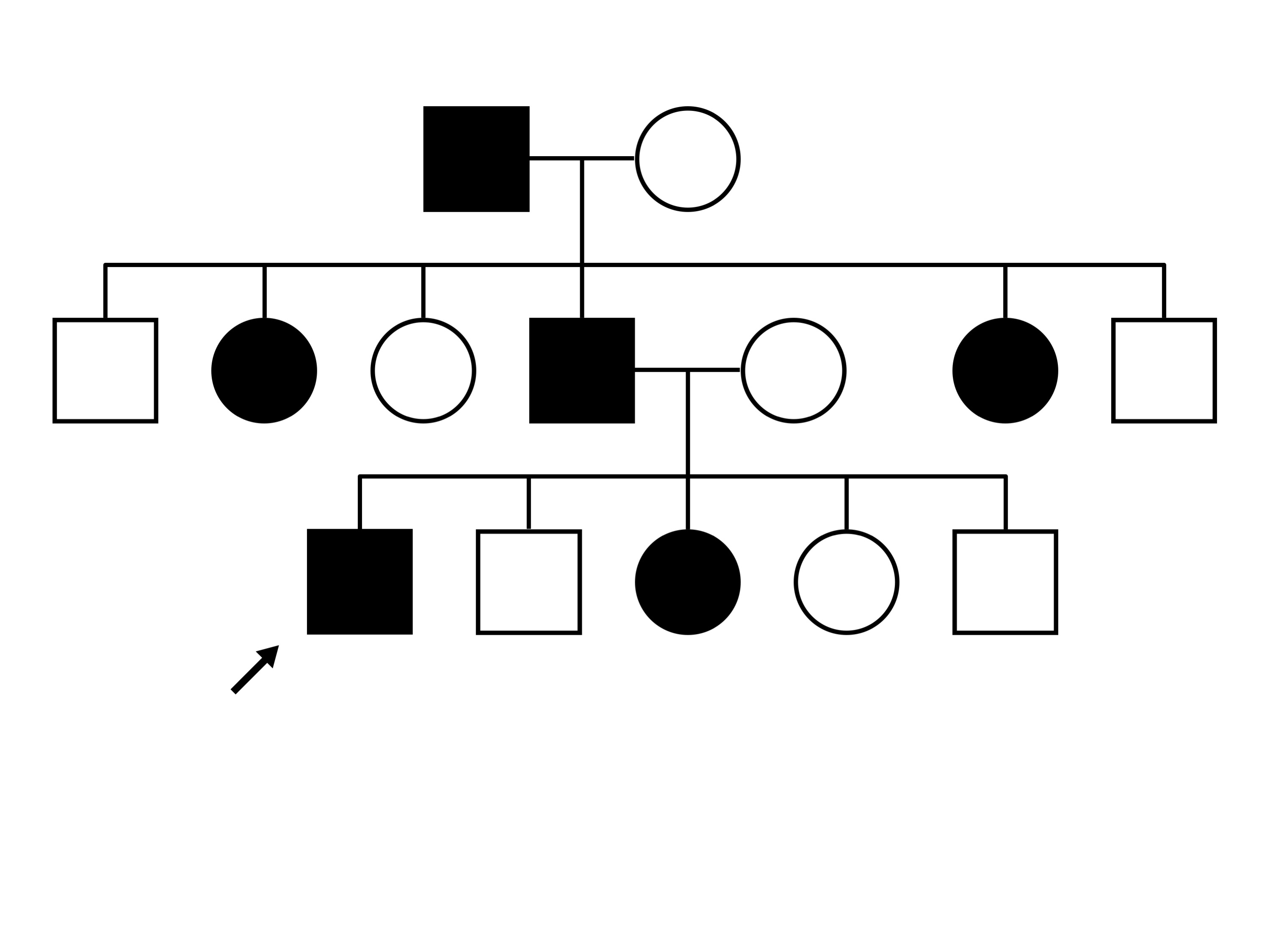
Pedigree
Teaching Points
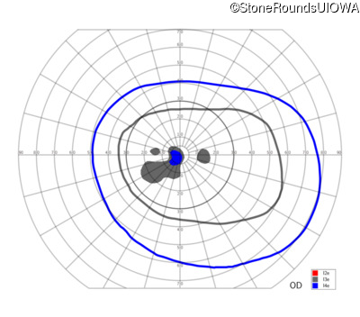
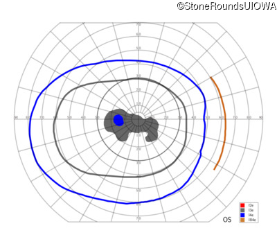
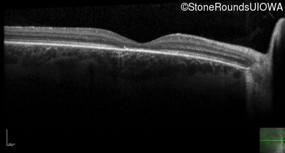
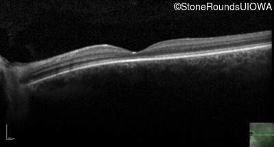
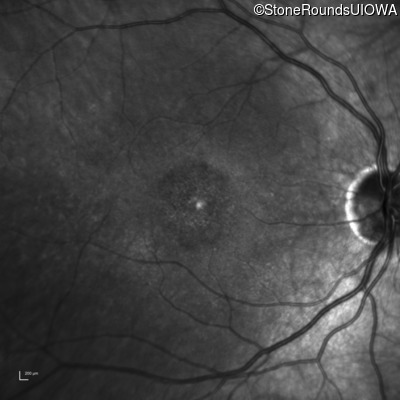
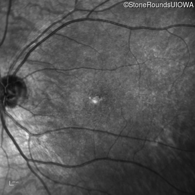
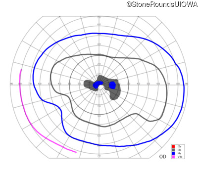
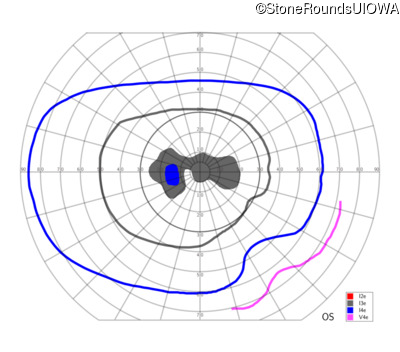
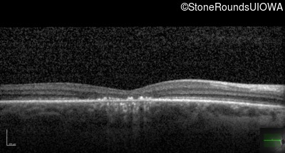
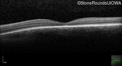
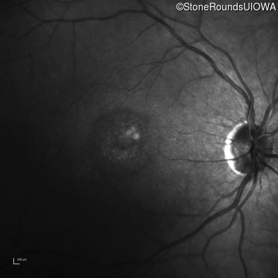
The clinical features supporting the diagnosis of autosomal dominant cone dystrophy in this patient include: reduced acuity and photophobia as his earliest symptoms; a bull's eye appearance of the fovea with a relatively normal fundus appearance more anteriorly; loss of foveal photoreceptors on OCT; global depression of the Goldmann visual field (complete loss of the I2e isopter); and, a three generation family history of similarly affected individuals with some male to male transmission.
| Age at visit: 30 years |

Diagnosis & molecular findings
| Disease | Gene | Allele 1 variant(s) | Allele 2 variant(s) | Inheritance mode |
|---|---|---|---|---|
| AD Cone and Cone Rod Dystrophy | GUCA1A | Glu155Gly GAG>GGG | AD |
Disease:
Gene:
Allele 1:
Glu155Gly GAG>GGG
Allele 2:
Inheritance:
AD