SR697
AR Retinitis Pigmentosa (IA1aiii)
Male
Male
SR697
AR Retinitis Pigmentosa (IA1aiii)
Male
Male
Highlighted Images
| Age at visit: 64 years | OD | OS |
|---|---|
History
This 64 year old myopic man had a traumatic retinal detachment in the right eye successfully repaired with a scleral buckle at age 31. Fourteen years later (age 45) he began noticing some reduction in night vision and peripheral vision in both eyes. As a young child, his best corrected visual acuity was completely normal.
| Refraction OD: | +2.00 +2.25 x 75 (aphakic, scleral buckle) |
|---|---|
| Refraction OS: | -7.00 +0.50 x 75 |
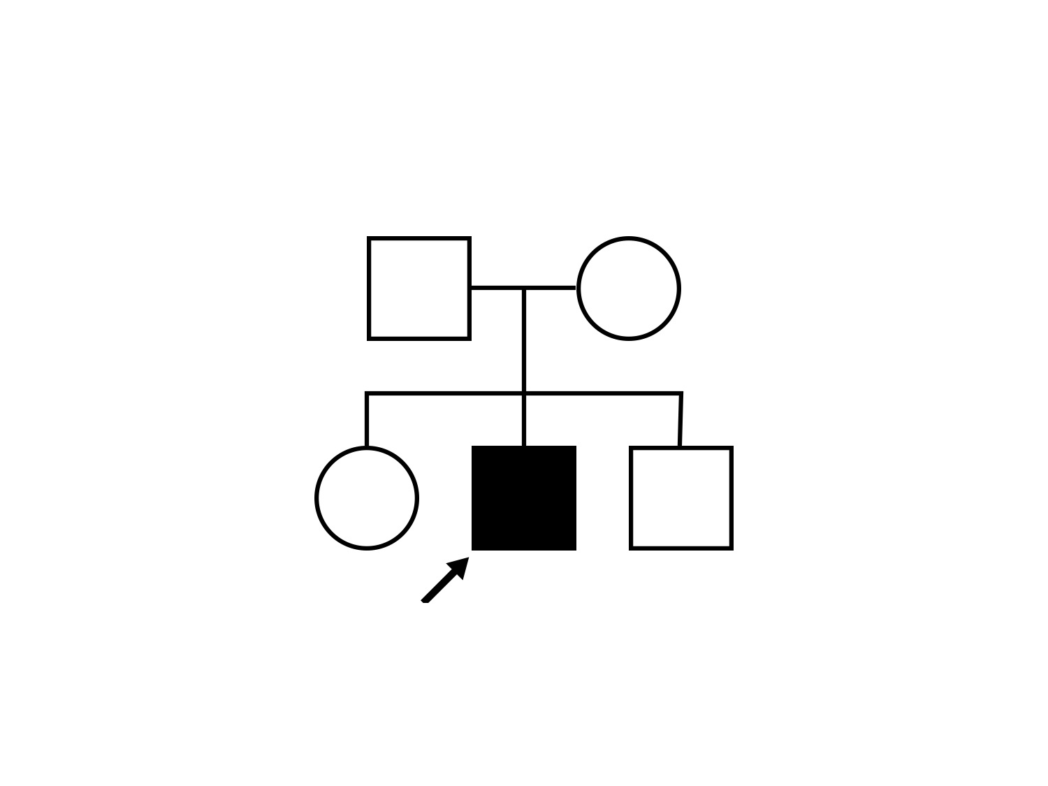
Pedigree
Teaching Points
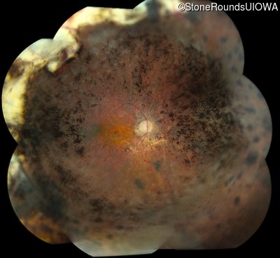
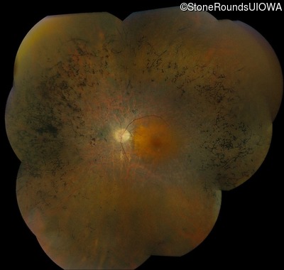
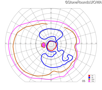
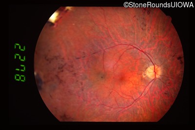
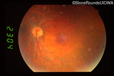
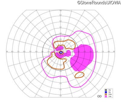
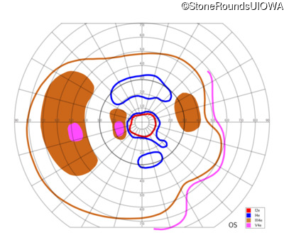
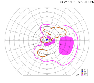
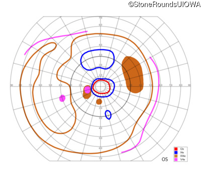
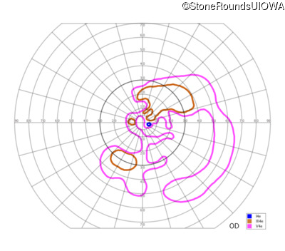
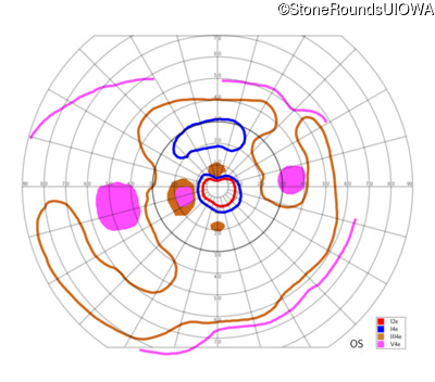
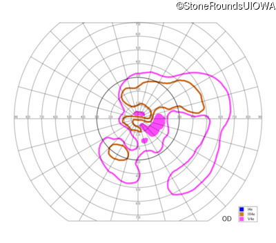
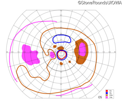
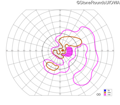
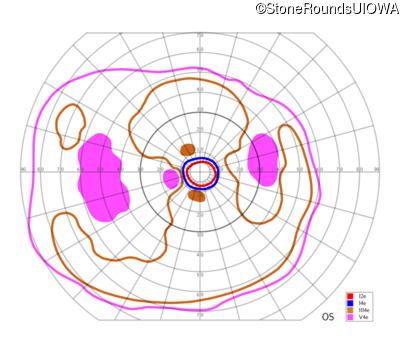
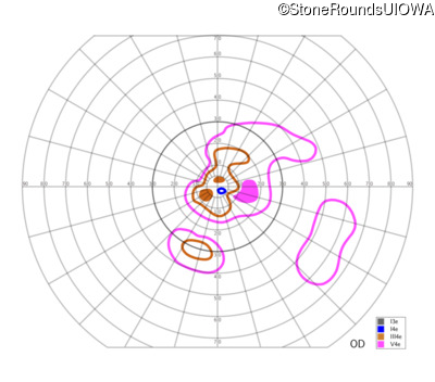
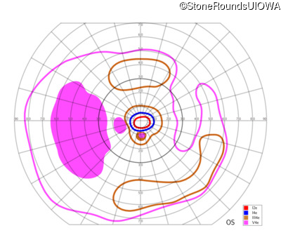
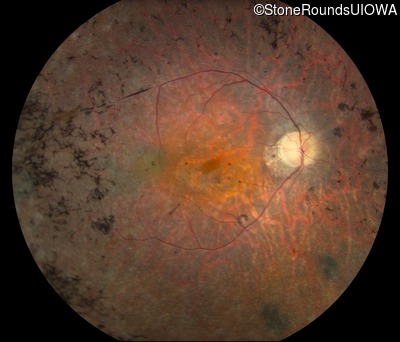
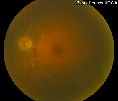
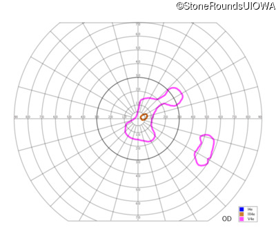
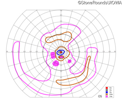
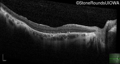
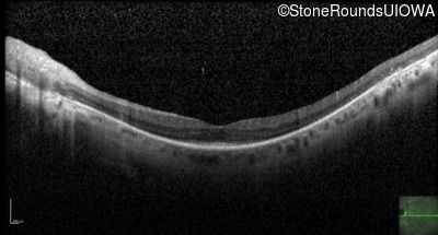
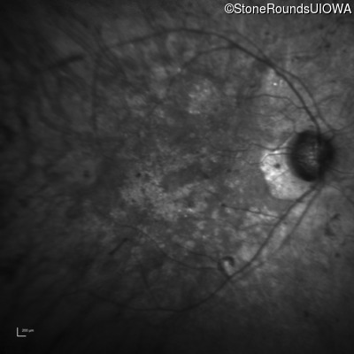
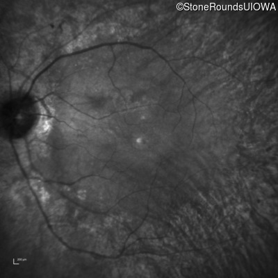
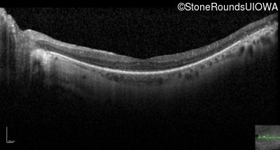
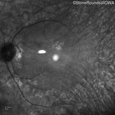
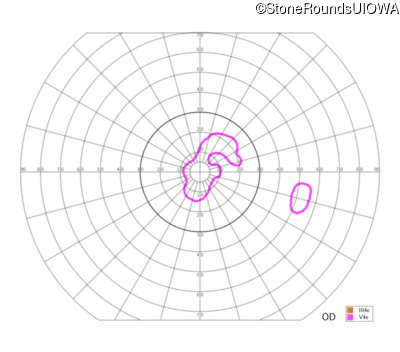
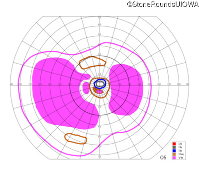
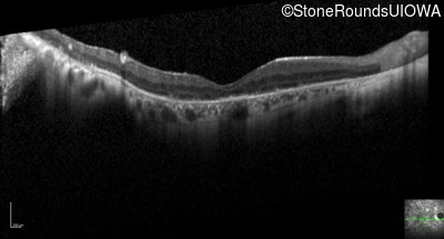
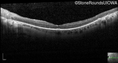
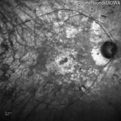
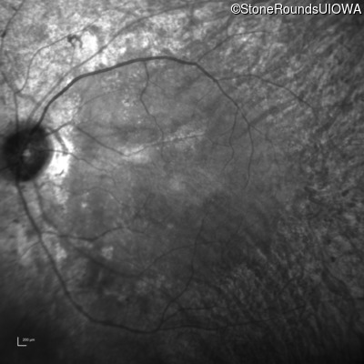
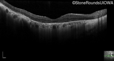
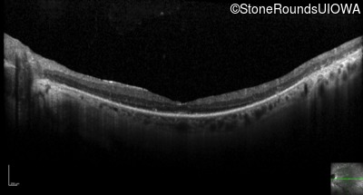
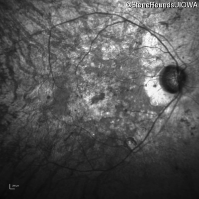
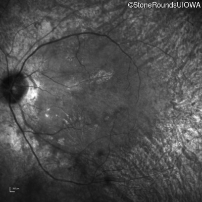
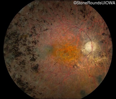
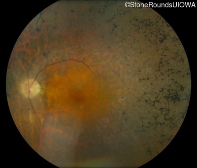
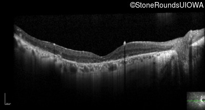
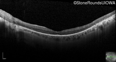
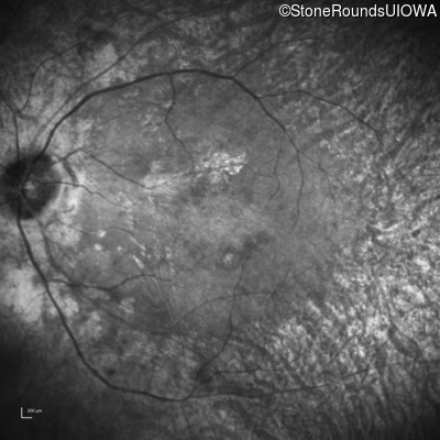
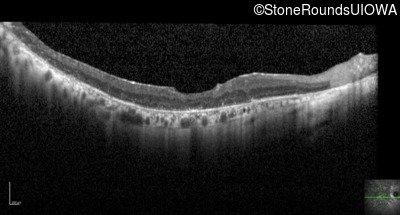
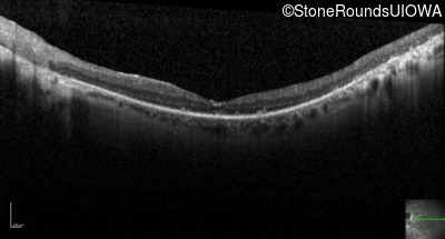
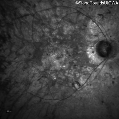
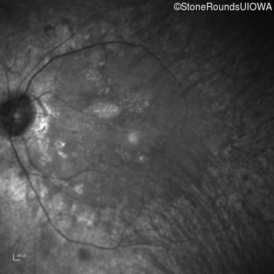
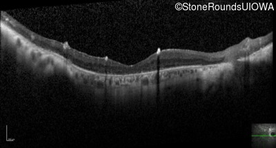
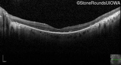
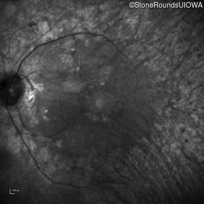
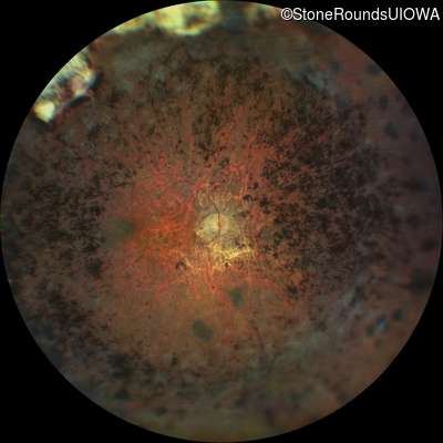
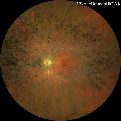
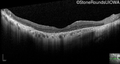
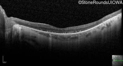
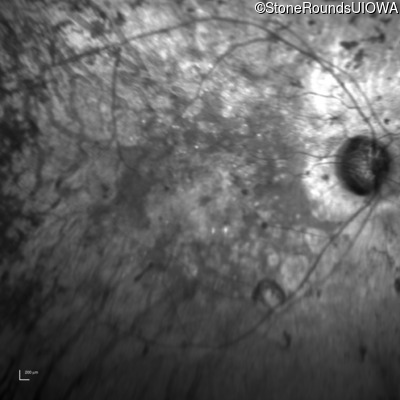
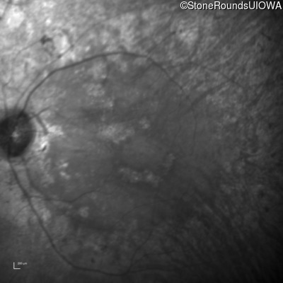
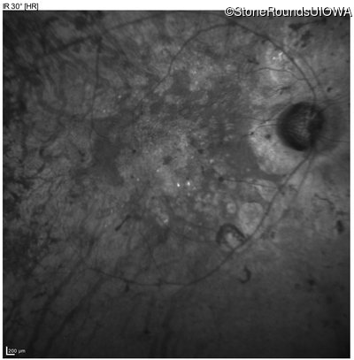
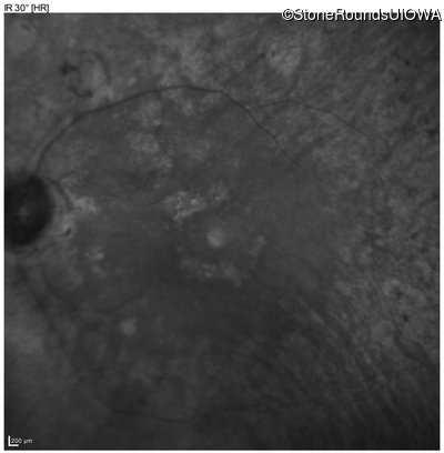
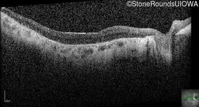
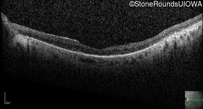
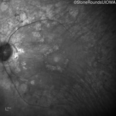
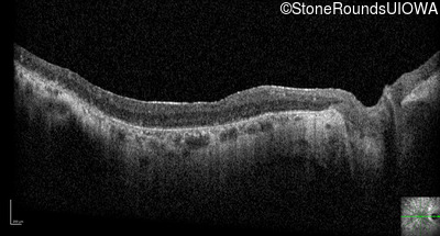
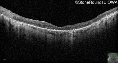
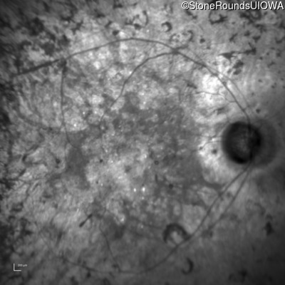
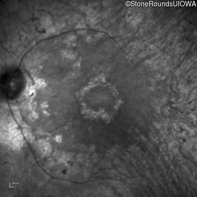
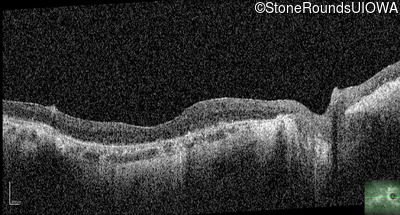
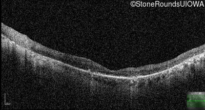
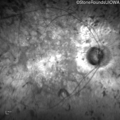
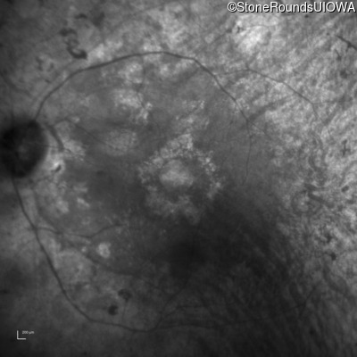
The clinical features supporting the diagnosis of autosomal recessive retinitis pigmentosa in this patient include: night blindness and constricted visual fields as his earliest symptoms; bone-spicule-like pigmentation and narrowed arterioles on fundus examination; loss of outer retinal structures on OCT; and, normally sighted parents.
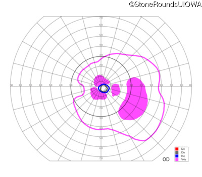
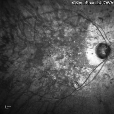
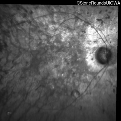
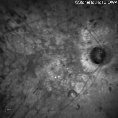
When one eye of a patient suspected to have an inherited eye disease has suffered some extensive surgery and/or trauma, it is best to focus initially on the symptoms of the unoperated/uninjured eye. Doing this, we find symptoms of night blindness and constricted peripheral vision both suggestive of adult onset rod-selective photoreceptor disease (retinitis pigmentosa). Fundus examination reveals very narrowed arterioles and bone-spicule-like pigmentation that are also compatible with this diagnosis. A ring scotoma is also evident on Goldmann perimetry of the left eye (it may be easiest to appreciate this pattern on the field of the left eye performed at age 48). Optical coherence tomography (see especially the OS at age 49) reveals such extensive loss of the outer nuclear layer that the outer plexiform layer could be mistaken for the external limiting membrane 2 millimeters eccentric to fixation. At this age (49 years) the visible ellipsoid zone in the left eye is only 1 mm in diameter and yet the patient can still detect the V4e stimulus 70 degrees from fixation inferiorly (superior retina). The fact that so few photoreceptors are needed for quite useful photopic vision is encouraging for stem cell transplantation strategies. Comparison of the visual fields of the two eyes (e.g., at age 48) suggests that the stress on the photoreceptors associated with the retinal detachment and associated surgery accelerated the progress of the retinitis pigmentosa to some degree. It is also interesting to compare the ophthalmoscopic view of the left fundus through a 20/60 cataract (age 59) with the view after cataract surgery 20/20, age 65). The montage fundus photograph of the right eye at age 65 gives an excellent view of the indentation of the retina caused by the scleral buckle and the cryotherapy scars superiorly and temporally associated with the successful treatment of the giant retinal tear.
| Age at visit: 53 years |
| Age at visit: 58 years |
| OD | OS | ||
|---|---|---|---|
| OD | OS | ||
|---|---|---|---|
| OD | OS | ||
|---|---|---|---|
| Age at visit: 61 years |
| OD | OS | ||
|---|---|---|---|
| Age at visit: 62 years |
| OD | OS | ||
|---|---|---|---|
| Age at visit: 63 years |
| Age at visit: 64 years |
| OD | OS | ||
|---|---|---|---|
| OD | OS | ||
|---|---|---|---|
| OD | OS | ||
|---|---|---|---|
| Age at visit: 67 years |
| Age at visit: 68 years |
| OD | OS | ||
|---|---|---|---|
| OD | OS | ||
|---|---|---|---|
| Age at visit: 70 years |
| OD | OS | ||
|---|---|---|---|
| Age at visit: 71 years |
| OD | OS | ||
|---|---|---|---|
Diagnosis & molecular findings
| Disease | Gene | Allele 1 variant(s) | Allele 2 variant(s) | Inheritance mode |
|---|---|---|---|---|
| AR Retinitis Pigmentosa | USH2A | IVS40-2144 A>G | Arg4192His CGC>CAC | AR |