Case
SR61
Student Mode
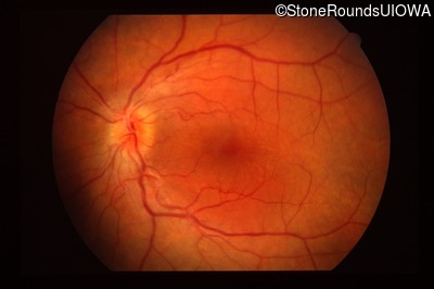
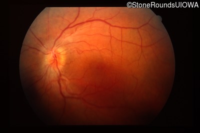
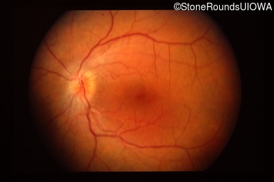
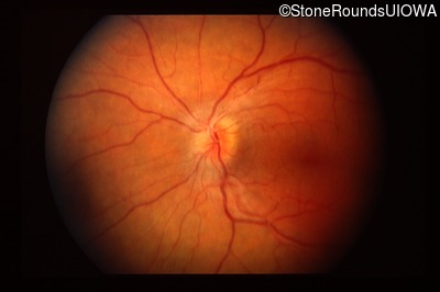
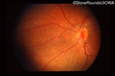
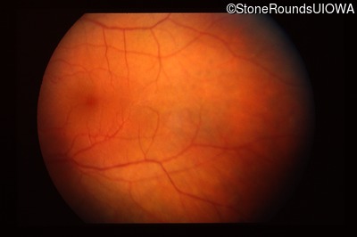
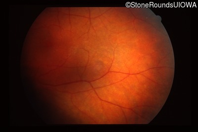
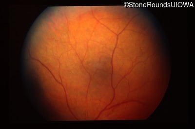
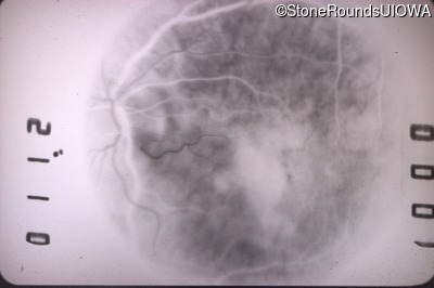
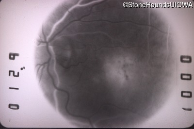
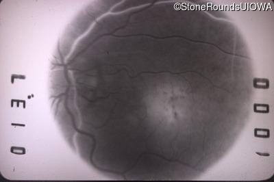
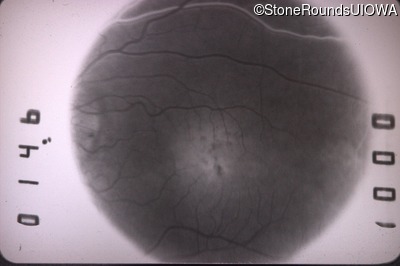
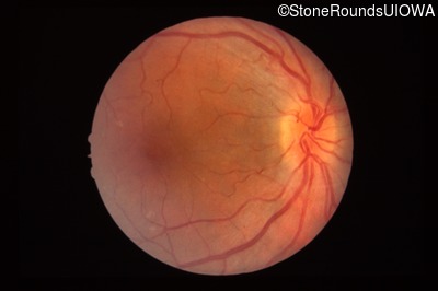
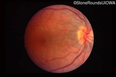
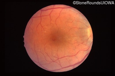
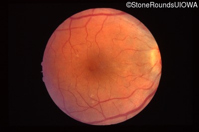
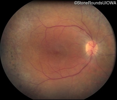
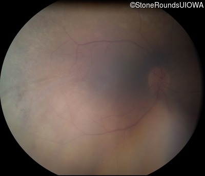
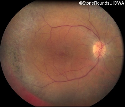
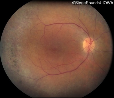
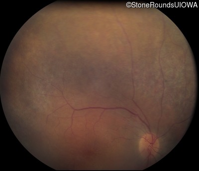
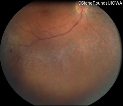
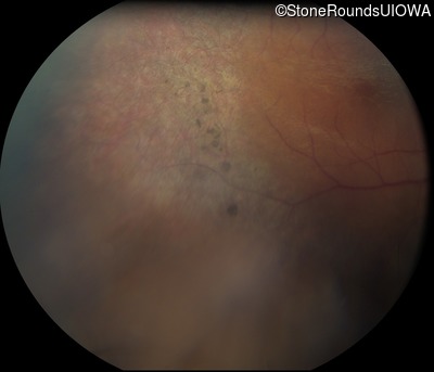
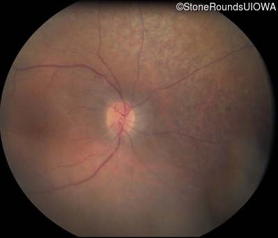
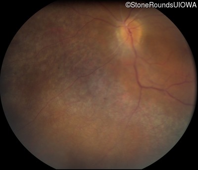
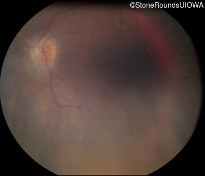
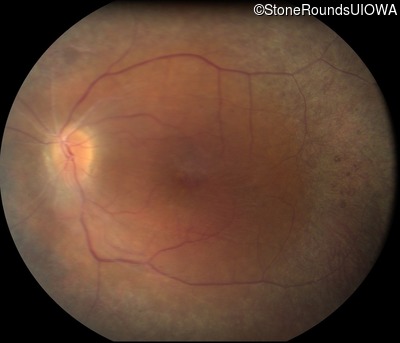
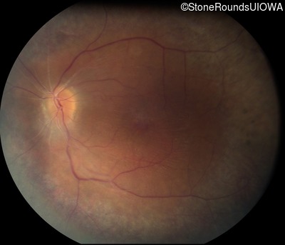
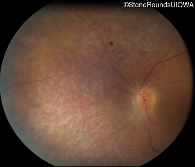
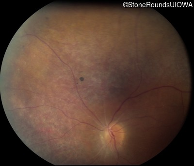
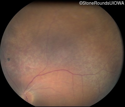
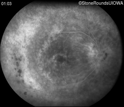
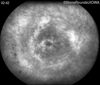
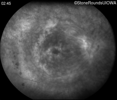
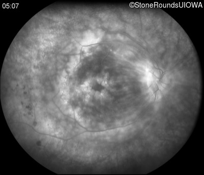
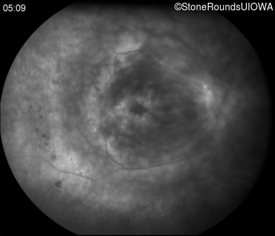
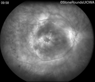
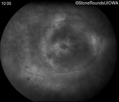
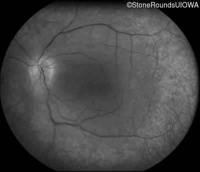
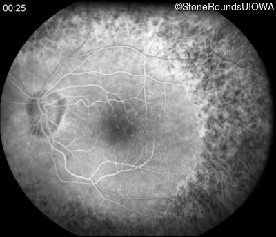
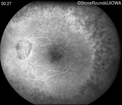
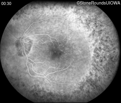
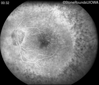
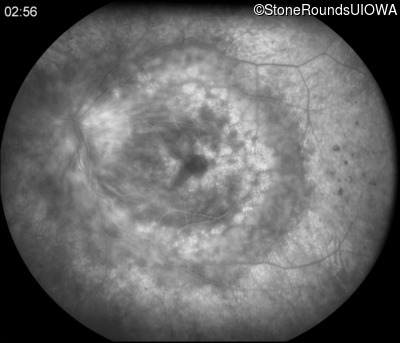
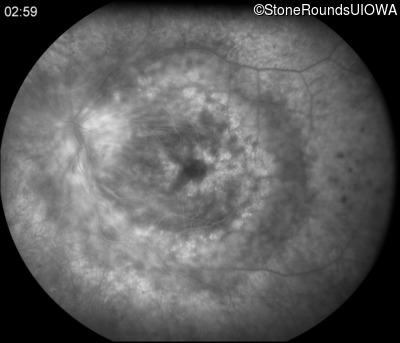
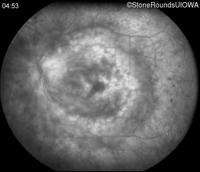
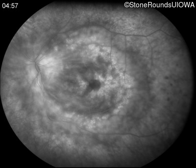
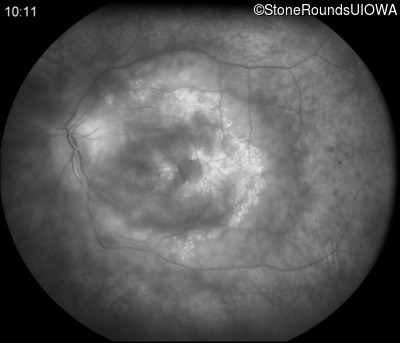
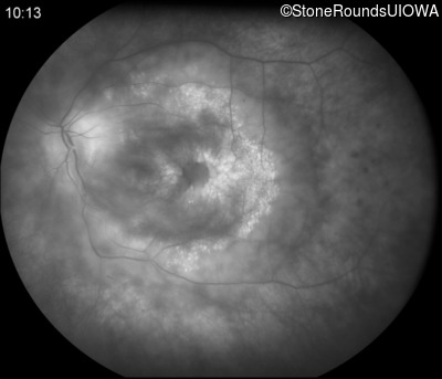
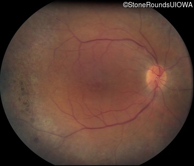
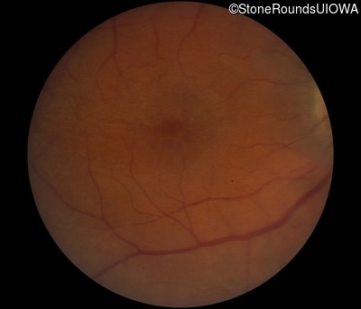
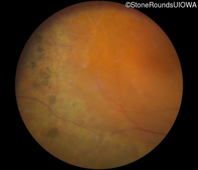
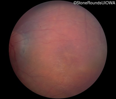
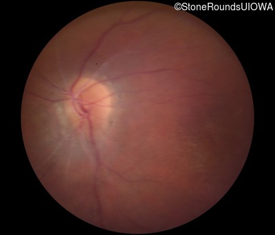
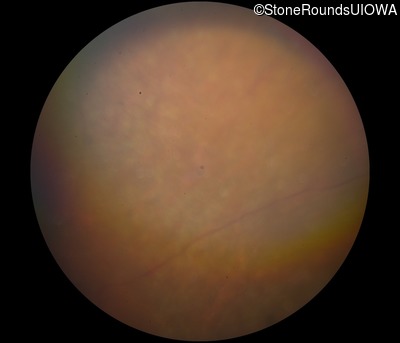
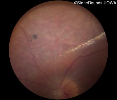
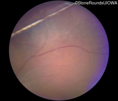
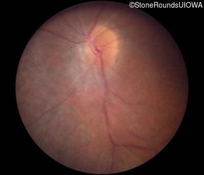
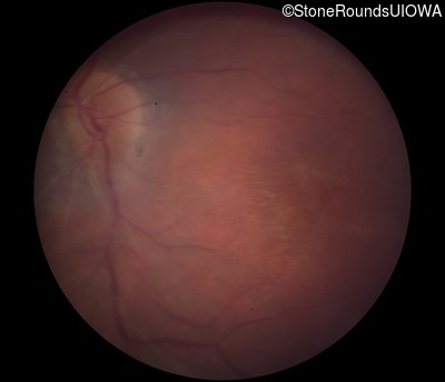
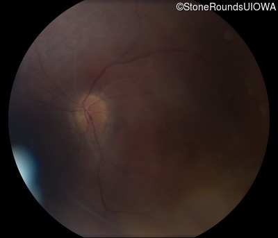
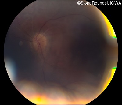
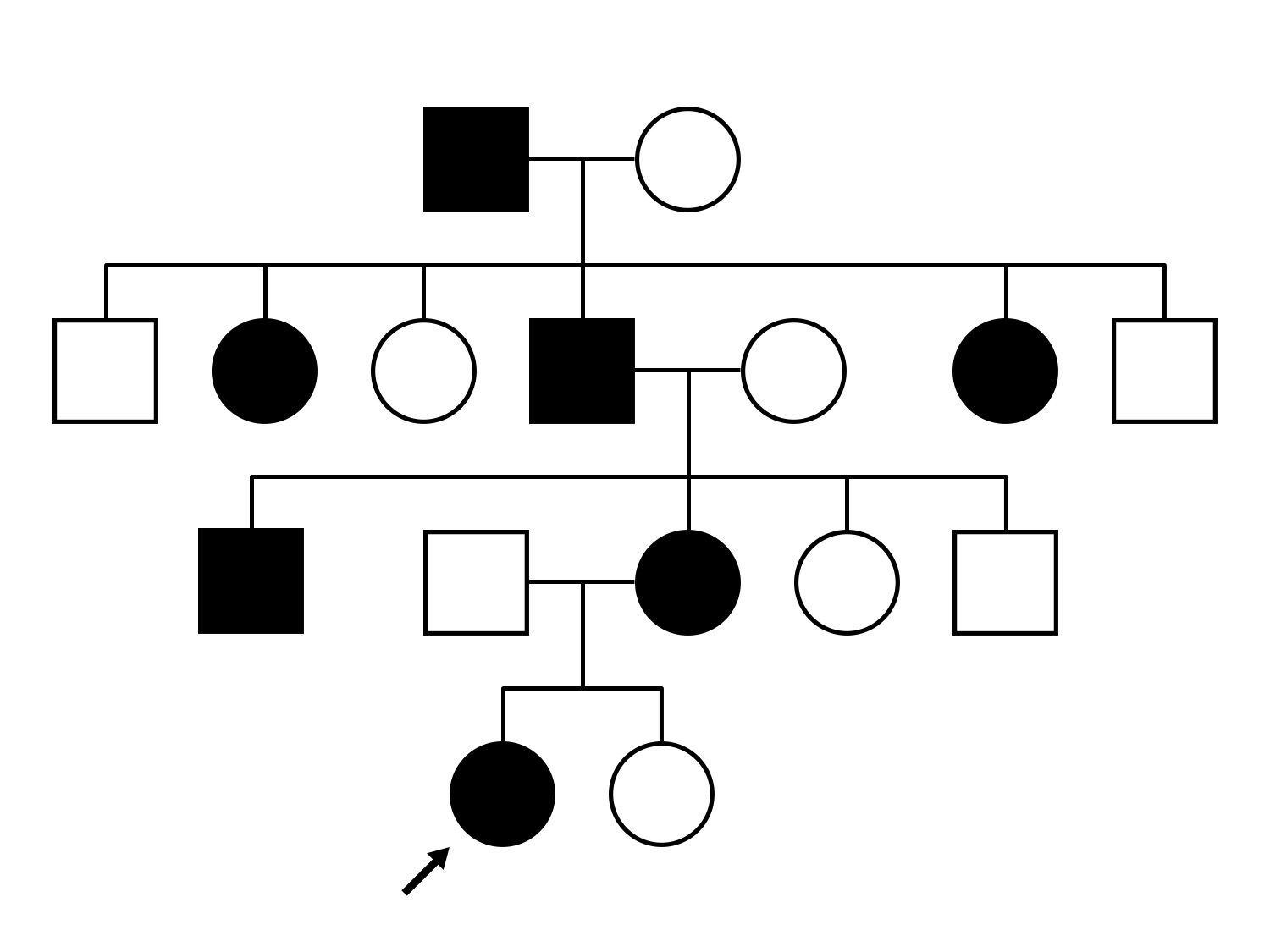
AD Neovascular Inflammatory Vitreoretinopathy (IIIE3)
Female
Female
Hidden
SR61
Student Mode
AD Neovascular Inflammatory Vitreoretinopathy (IIIE3)
Female
Female
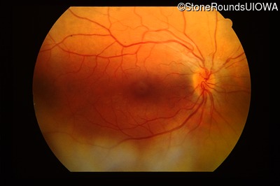
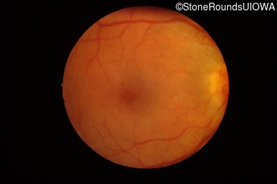
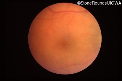
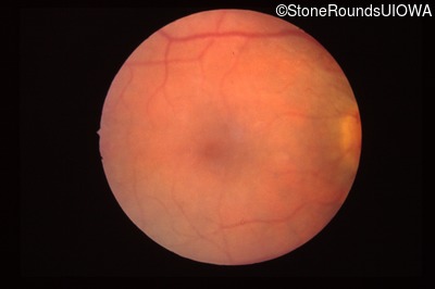
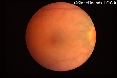
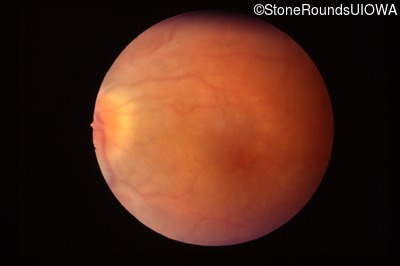
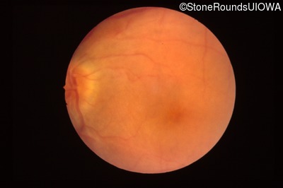
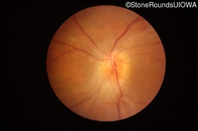
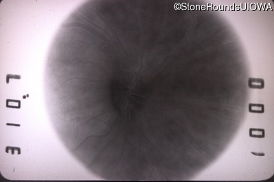
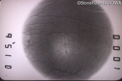
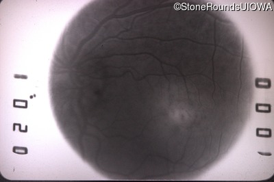
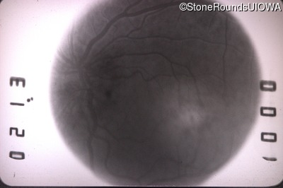
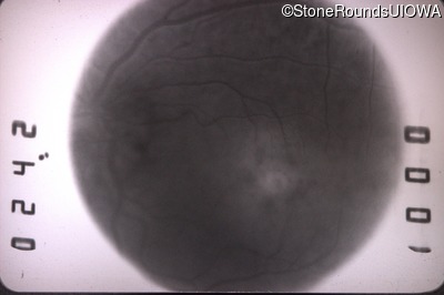
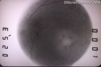
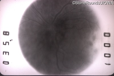
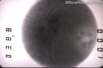
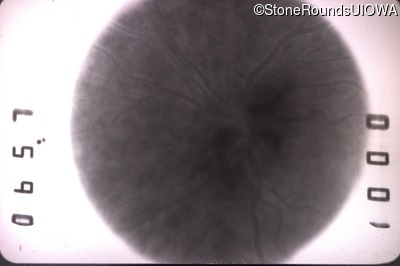
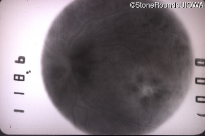
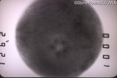
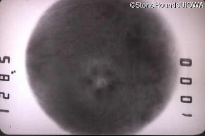
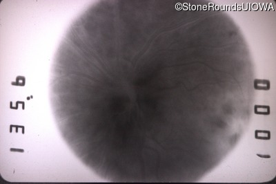
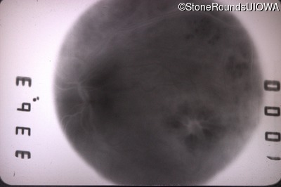
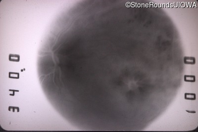
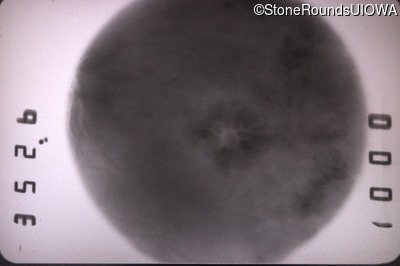
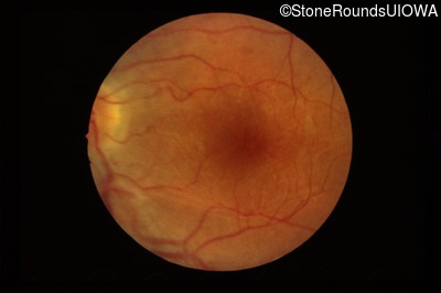
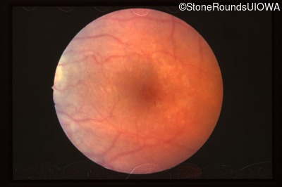
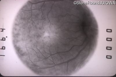
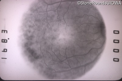
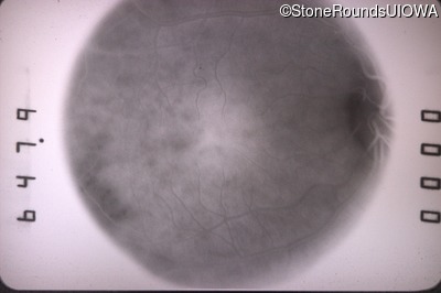
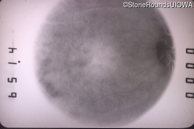
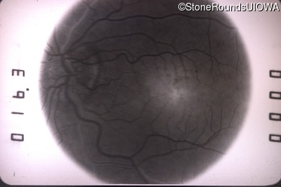
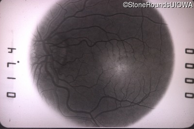
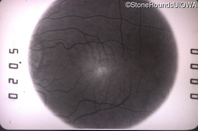
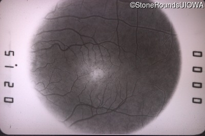
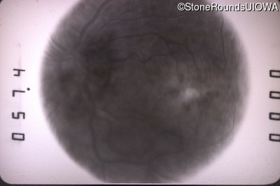
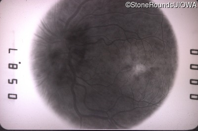
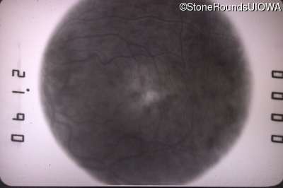
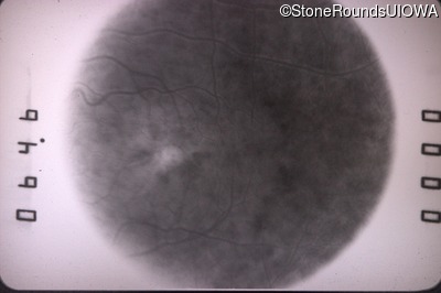
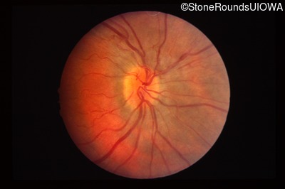
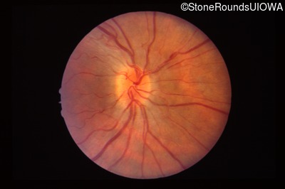
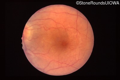
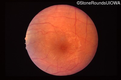
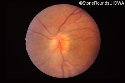
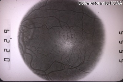
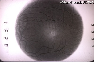
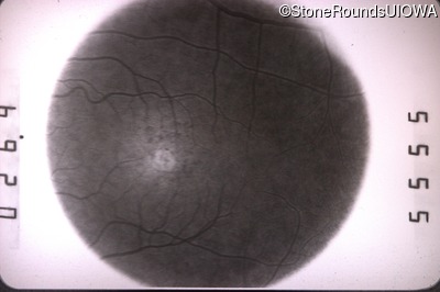
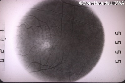
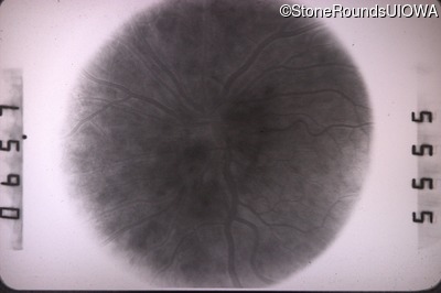
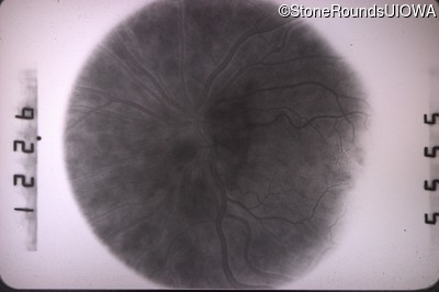
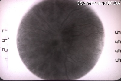
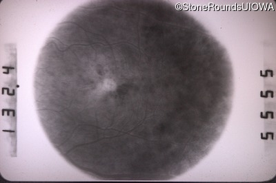
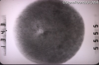
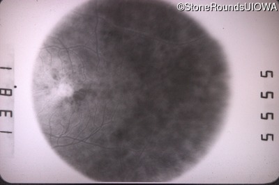
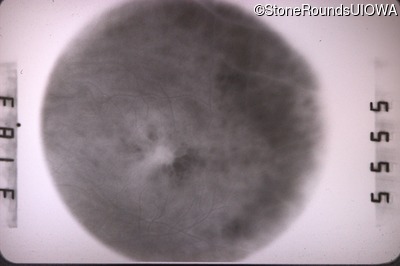
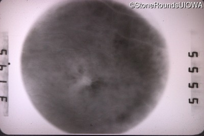
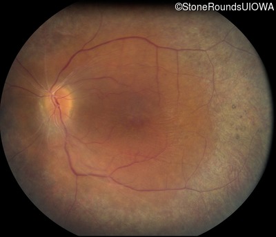
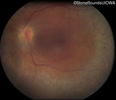
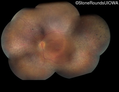
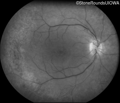
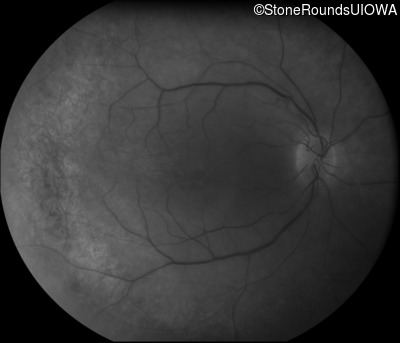
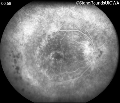
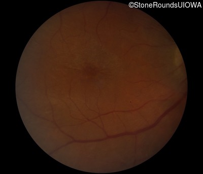
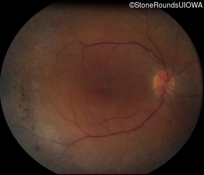
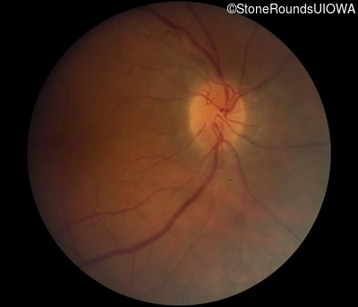
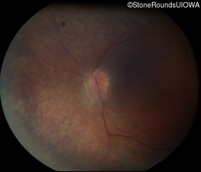
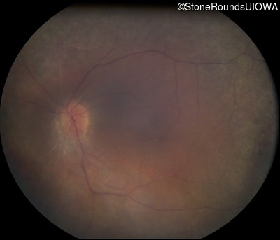
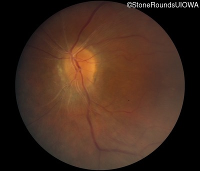
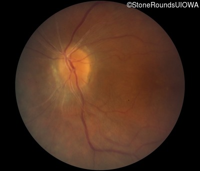
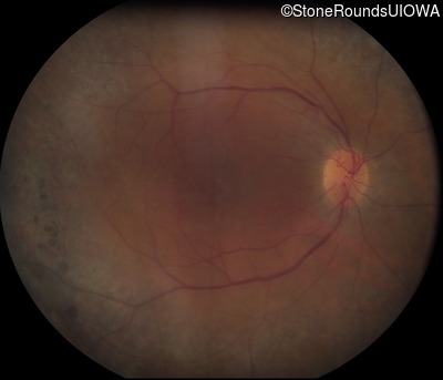
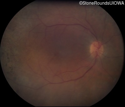
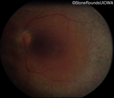
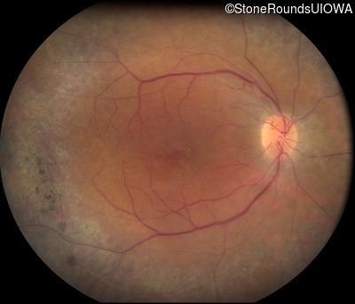
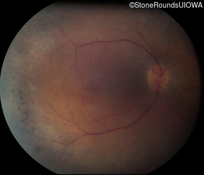
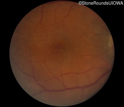
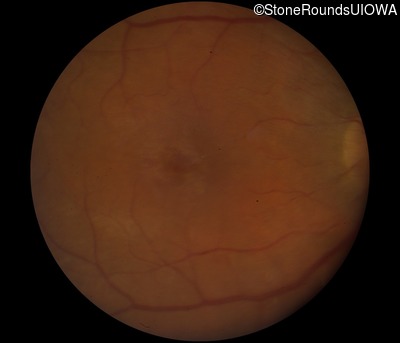
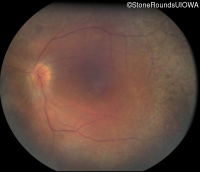
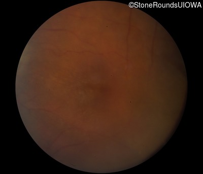
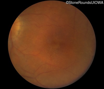
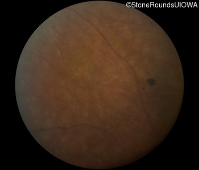
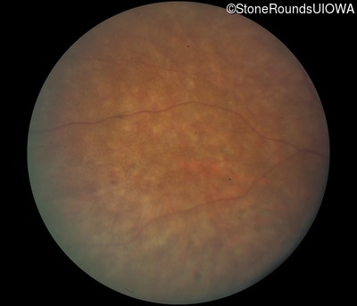
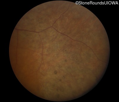
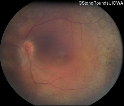
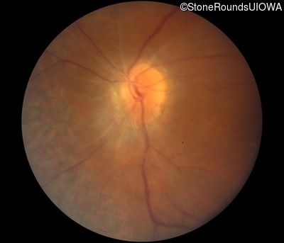
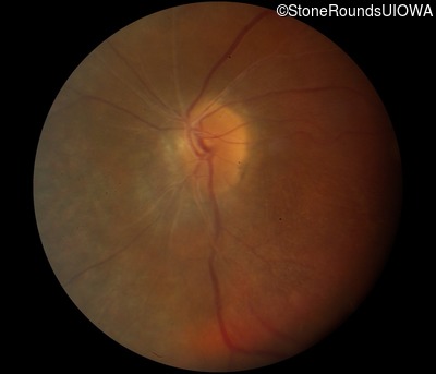
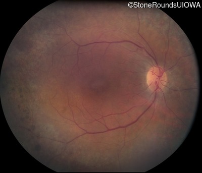
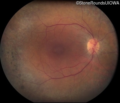
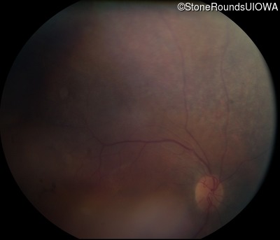
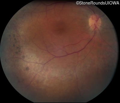
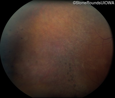
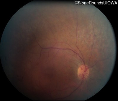
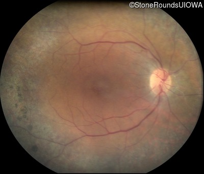
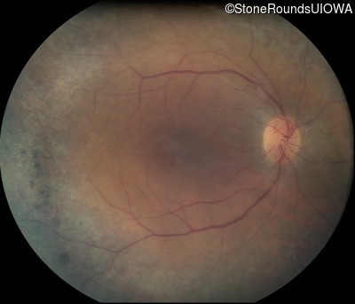
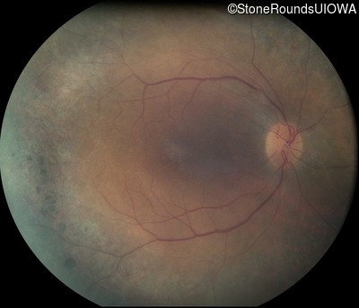
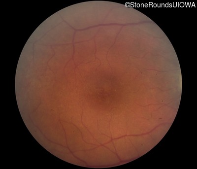
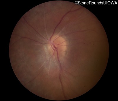
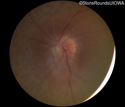
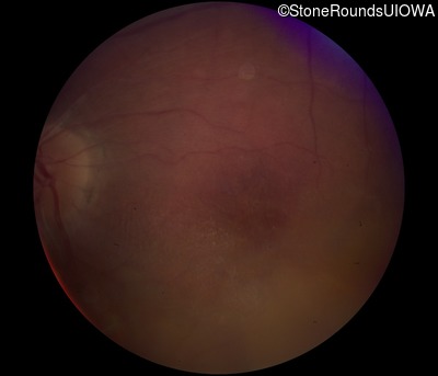
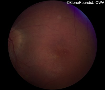
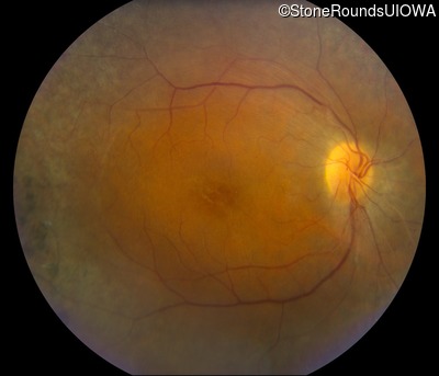
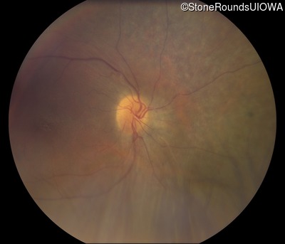
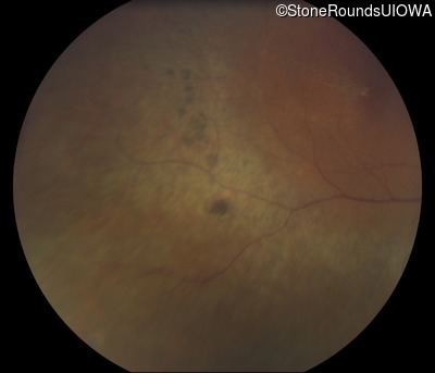
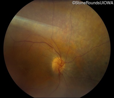
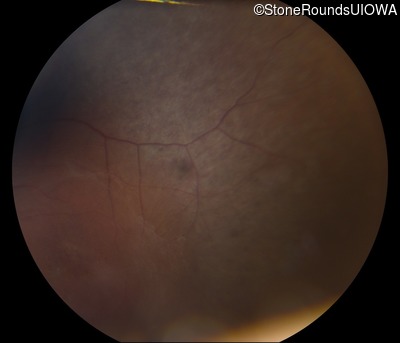
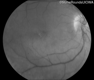
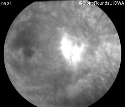
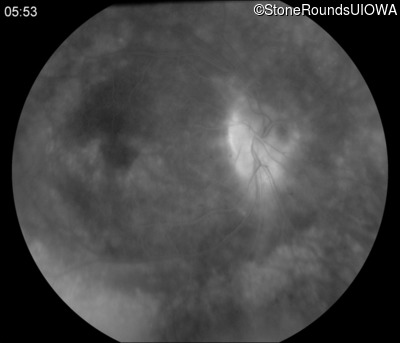
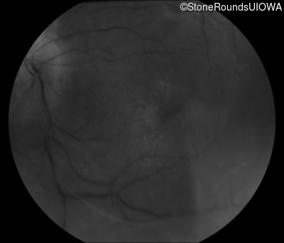
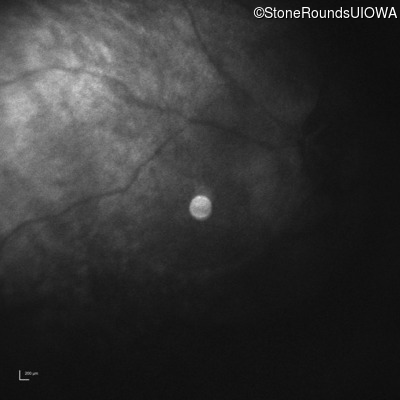
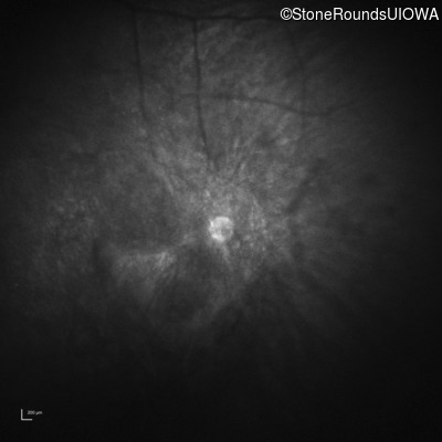
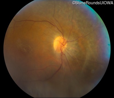
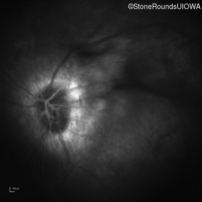
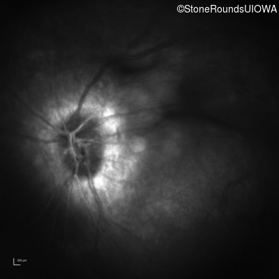
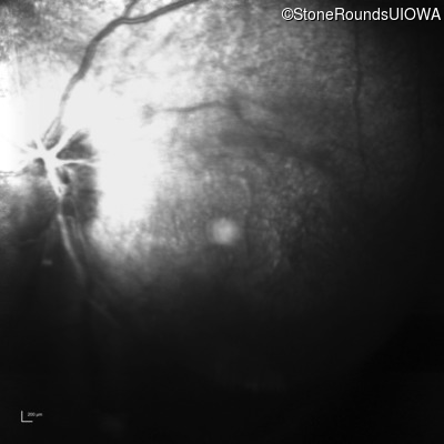
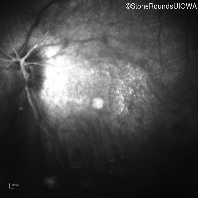
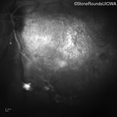
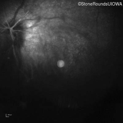
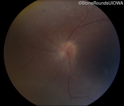
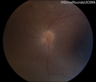
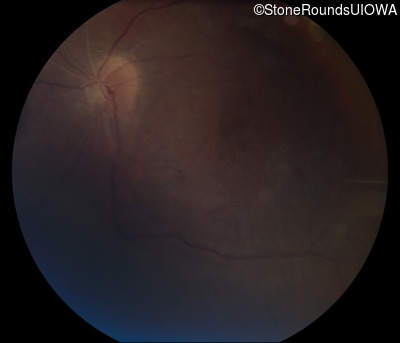
Visit at age: 23 years
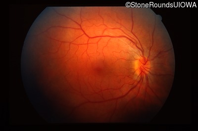
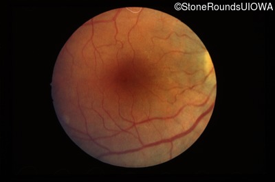
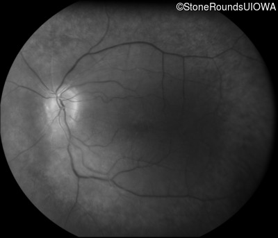
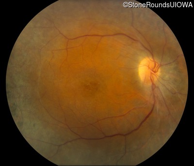
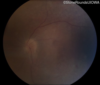
Fundus Photography - Right - 20/40
Exemplar
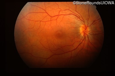
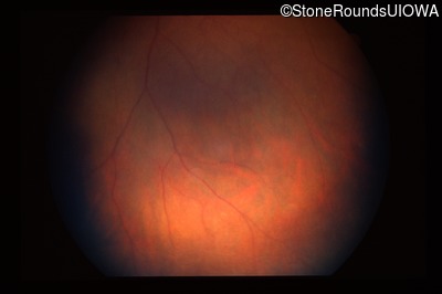
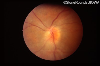
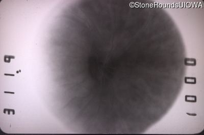
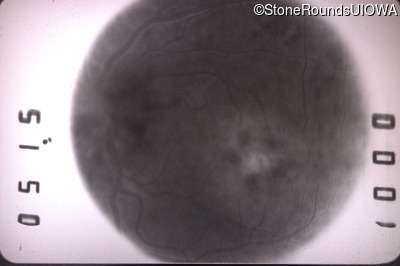
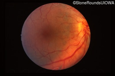
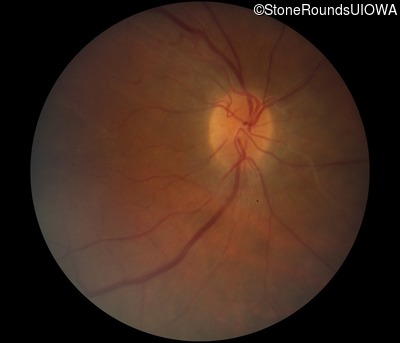
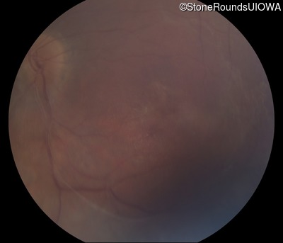
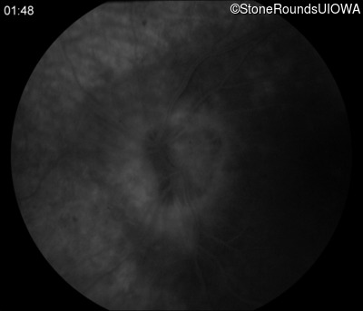
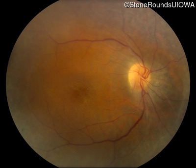
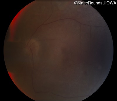
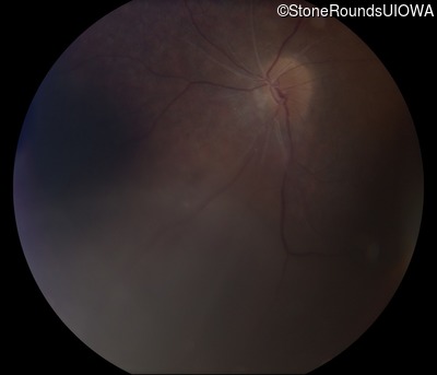
Fundus Photography - Left - 20/30
Exemplar
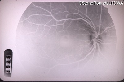
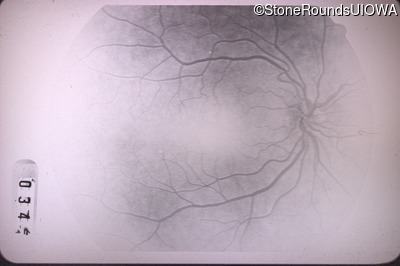
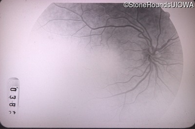
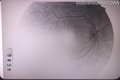
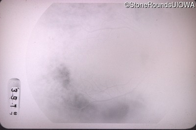
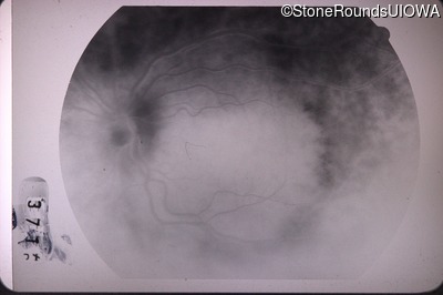
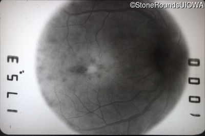
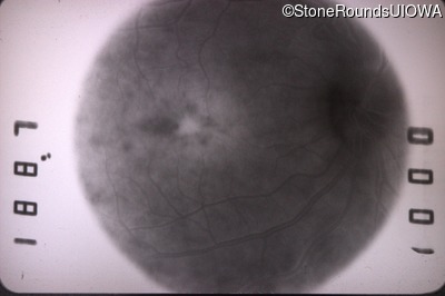
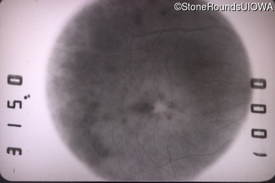
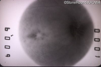
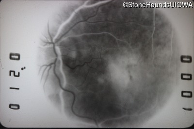
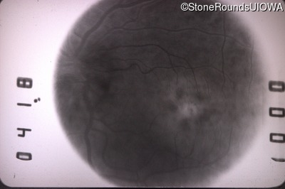
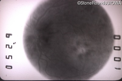
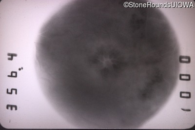
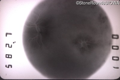
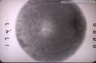
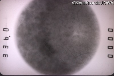
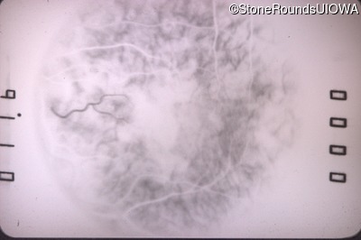
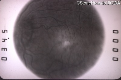
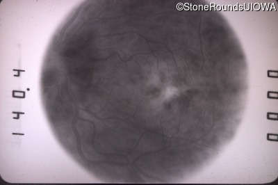
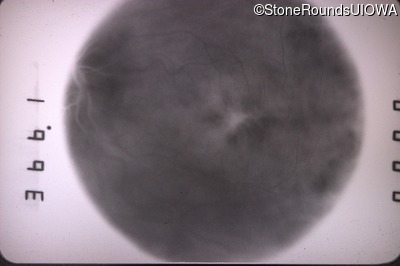
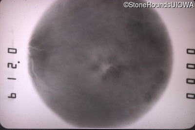
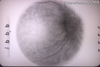
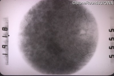
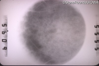
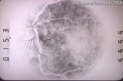
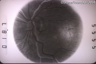
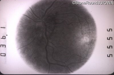
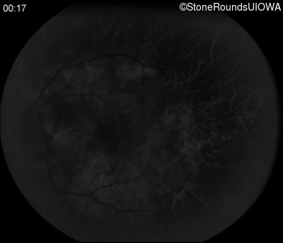
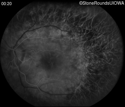
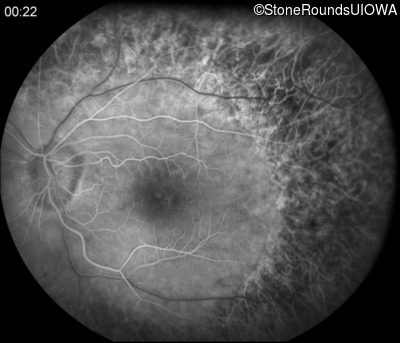
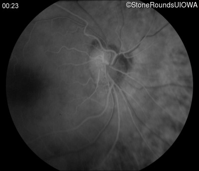
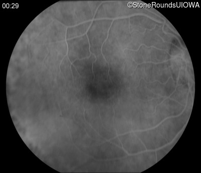
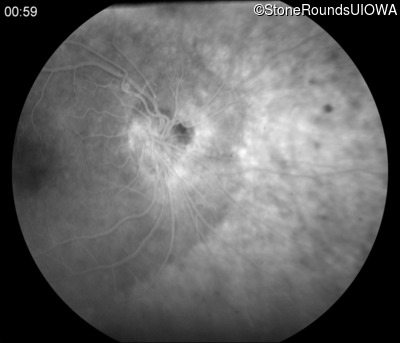
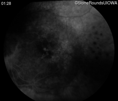
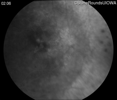
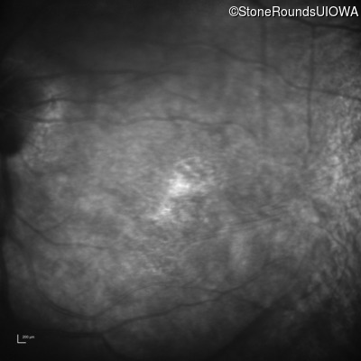
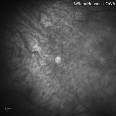
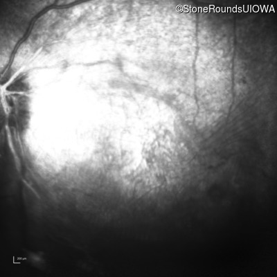
Fluorescein Angiography - Right - 20/40
Exemplar
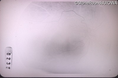
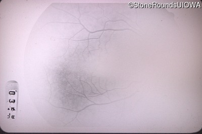
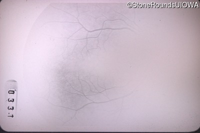
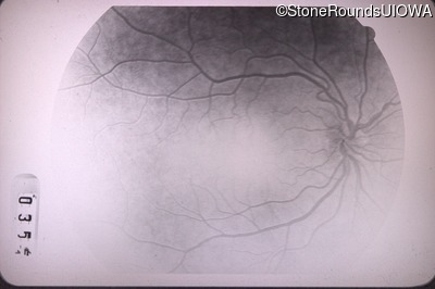
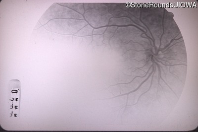
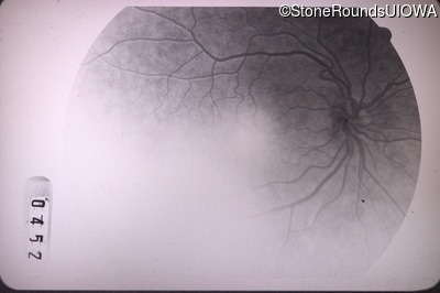
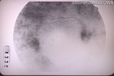
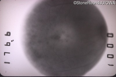
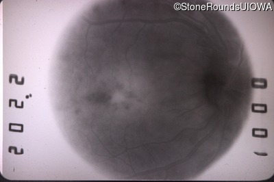
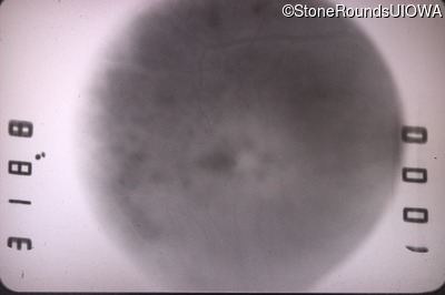
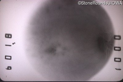
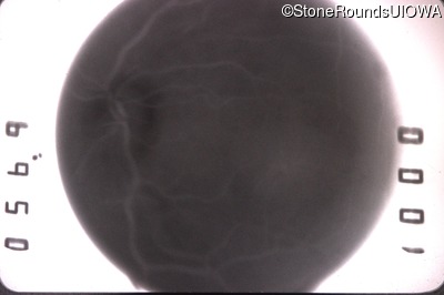
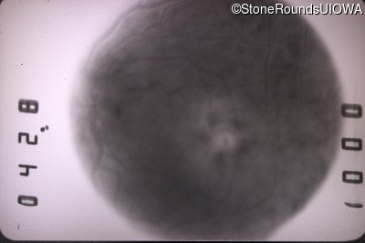
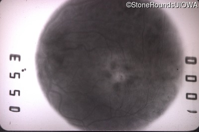
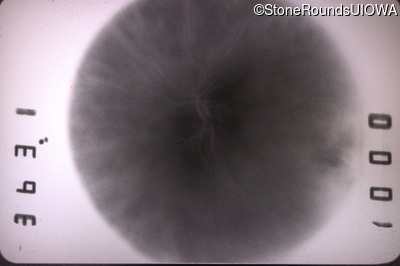
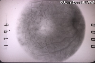
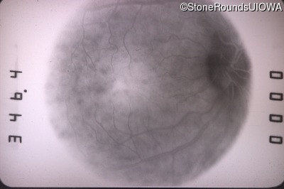
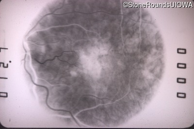
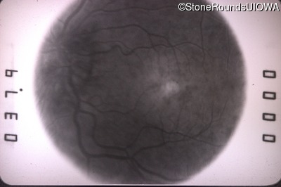
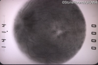
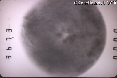
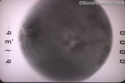
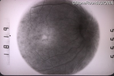
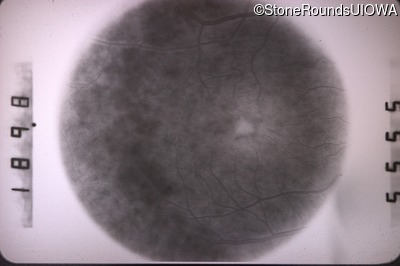
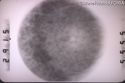
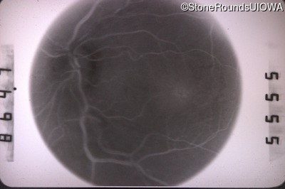
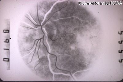
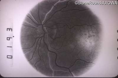
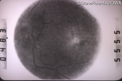
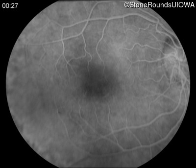
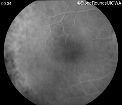
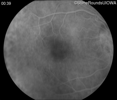
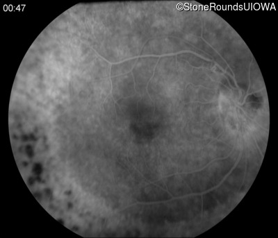
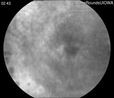
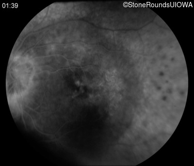
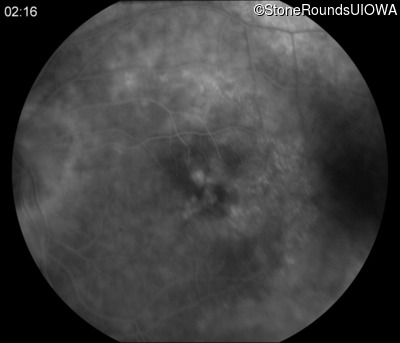
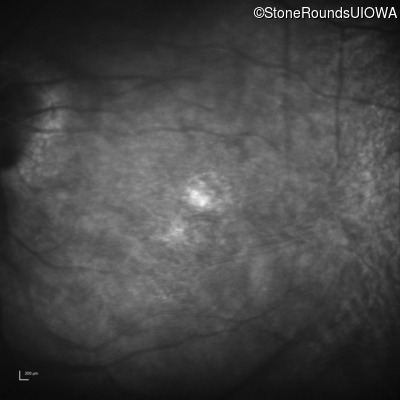
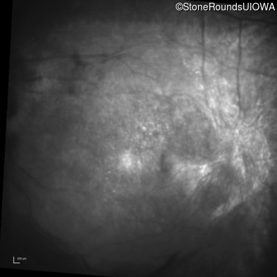
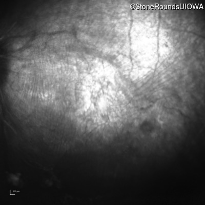
Fluorescein Angiography - Left - 20/30
Exemplar
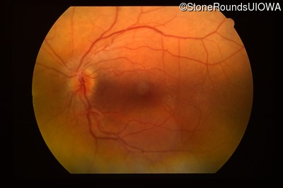
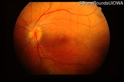
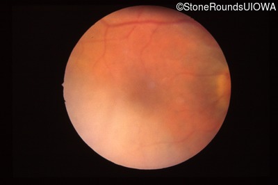
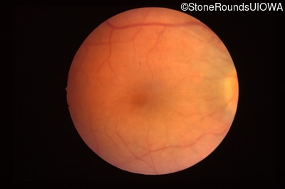
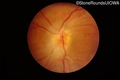
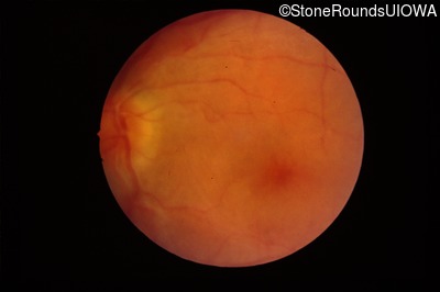
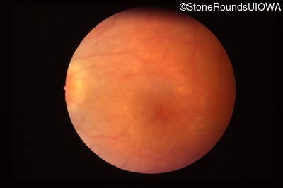
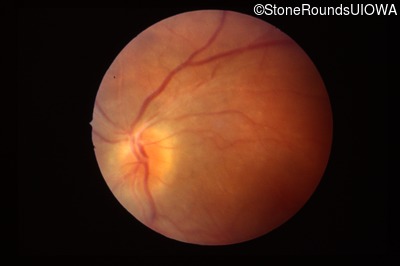
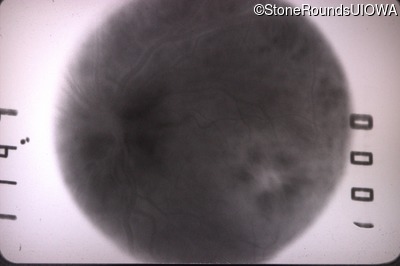
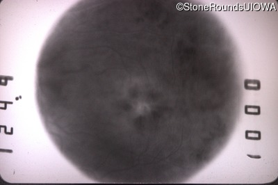
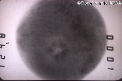
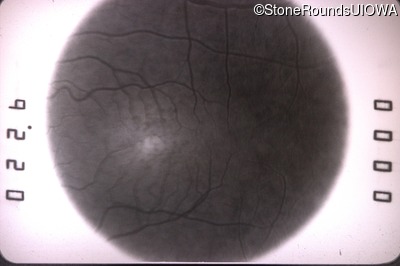
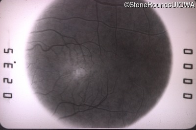
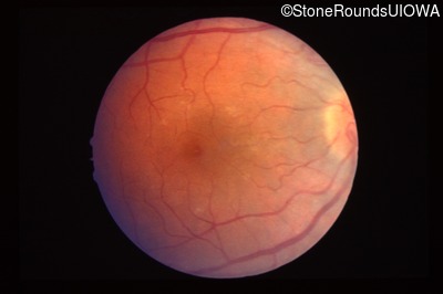
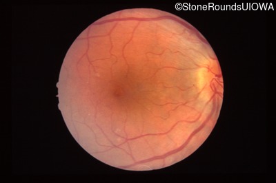
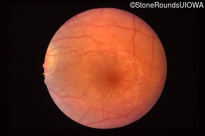
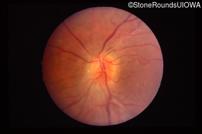
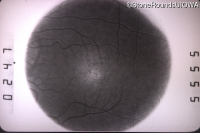
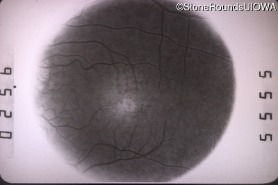
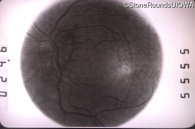
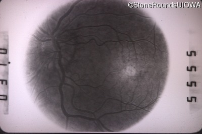
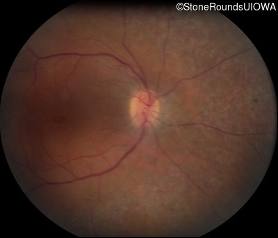
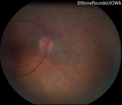
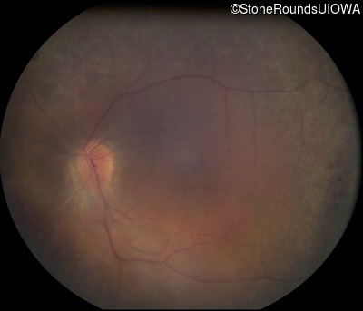
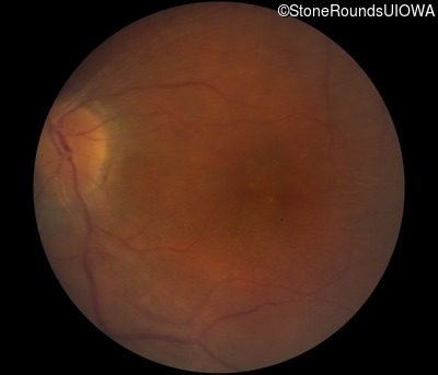
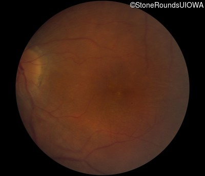
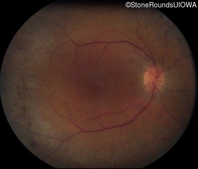
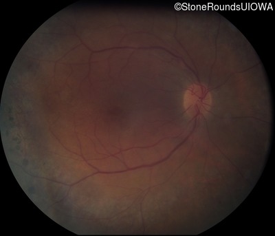
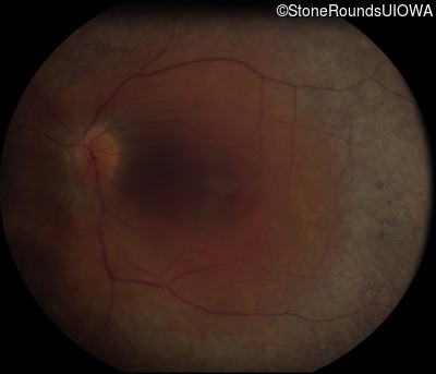
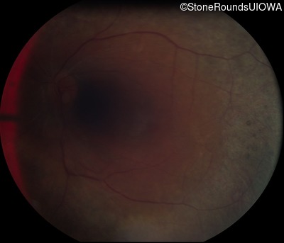
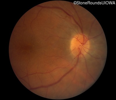
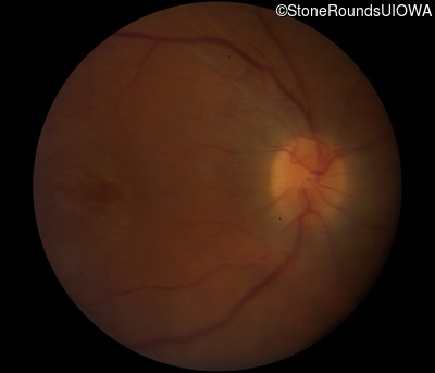
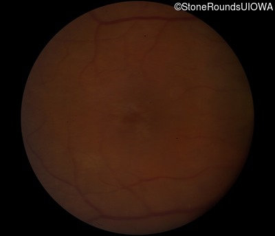
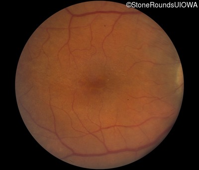
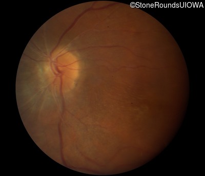
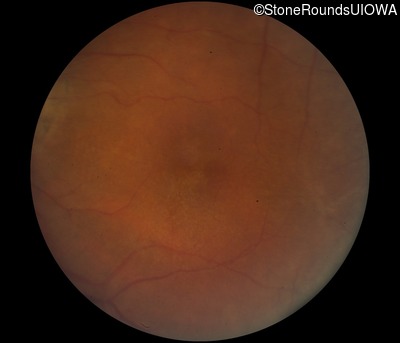
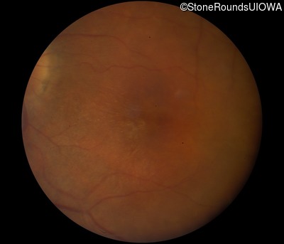
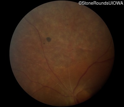
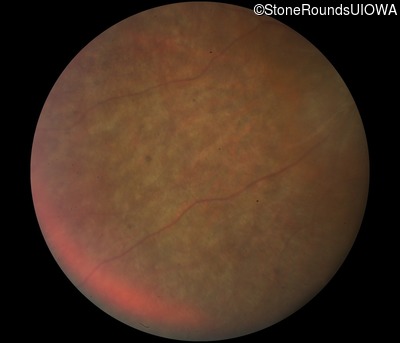
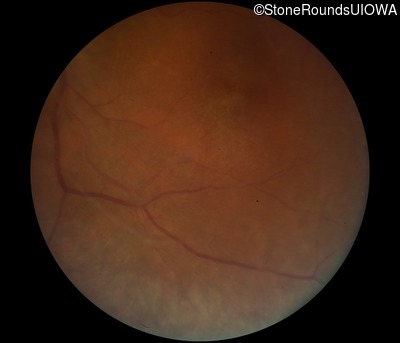
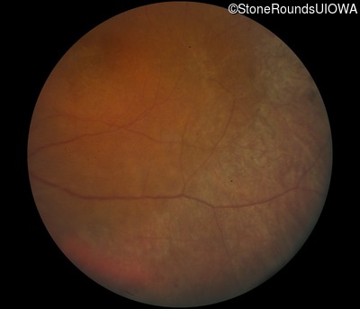
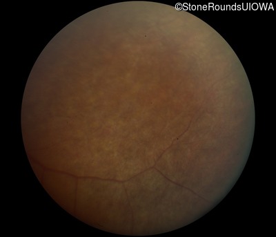
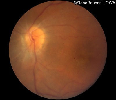
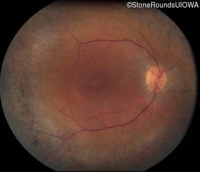
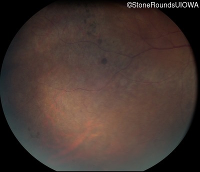
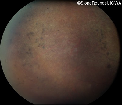
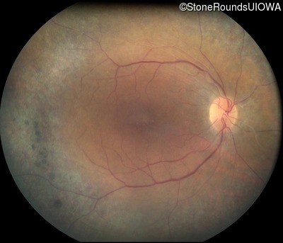
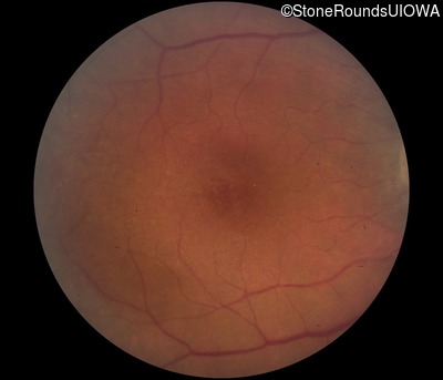
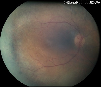
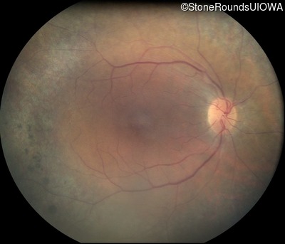
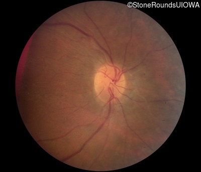
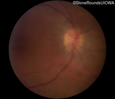
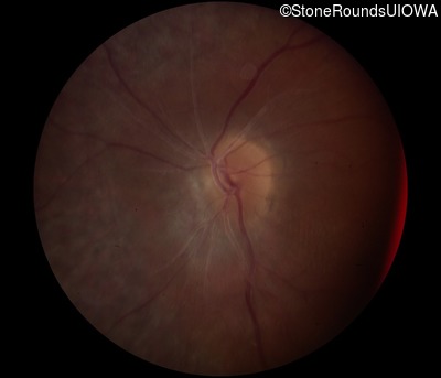
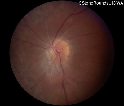
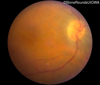
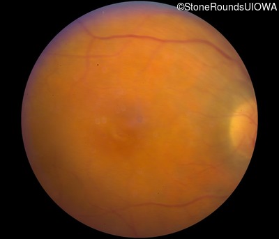
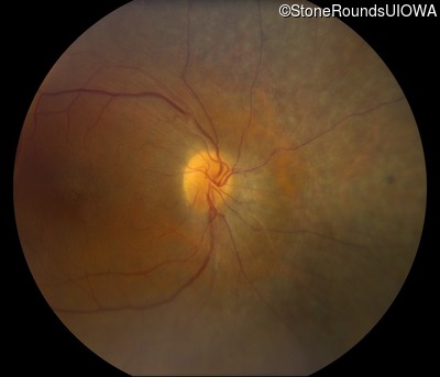
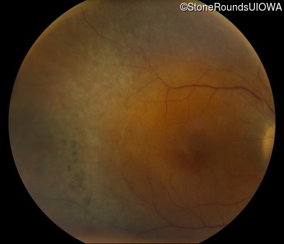
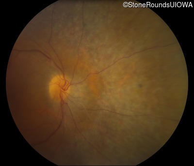
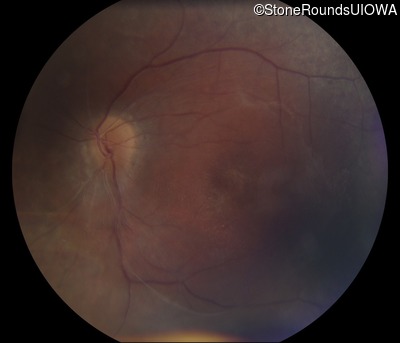
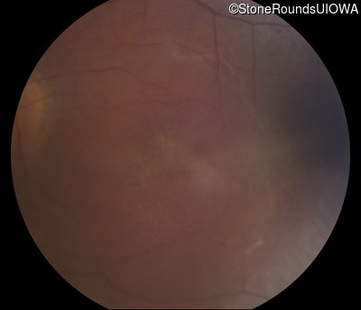
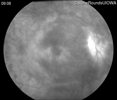
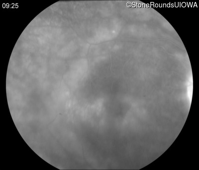
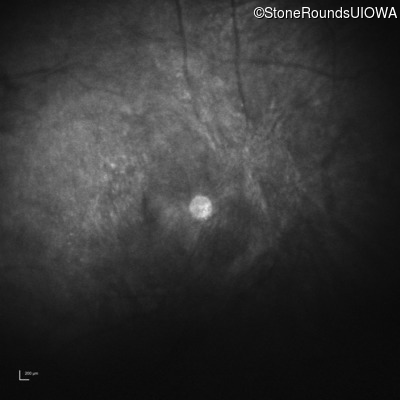
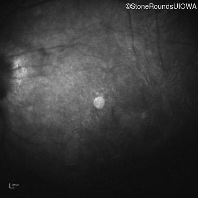
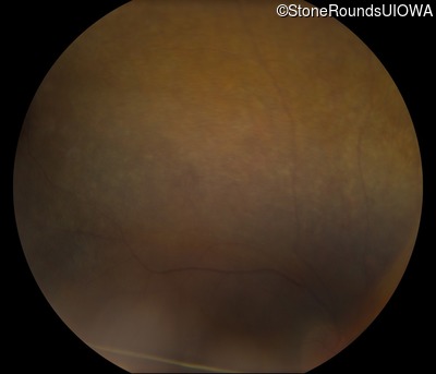
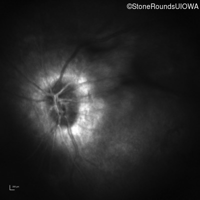
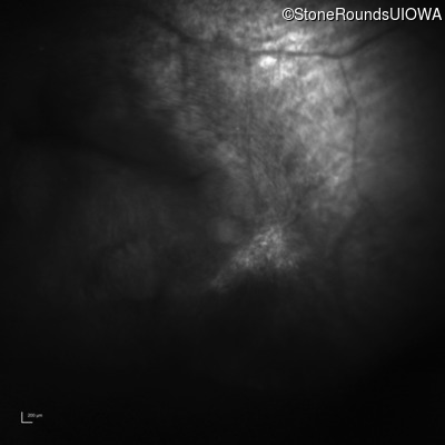
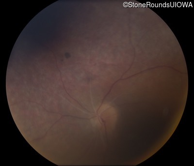
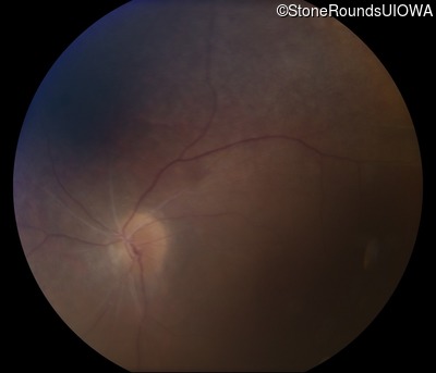
Visit at age: 25 years
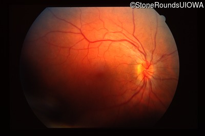
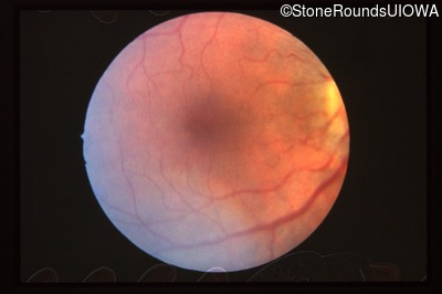
Fundus Photography - Right - 20/30
Exemplar
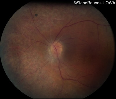
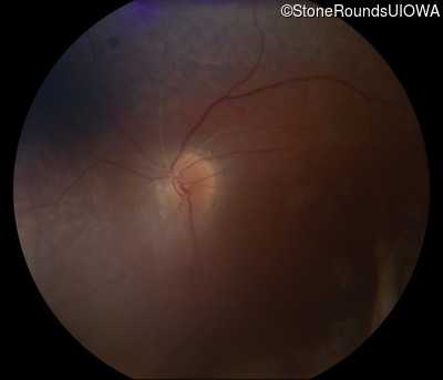
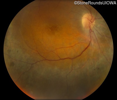
Fundus Photography - Left - 20/30
Exemplar
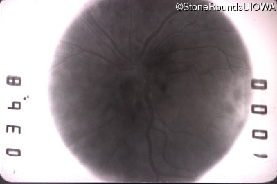
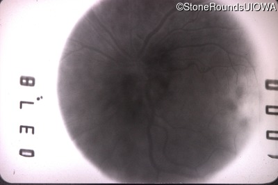
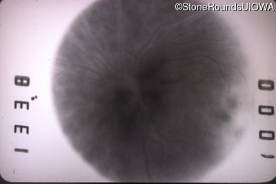
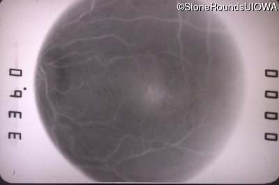
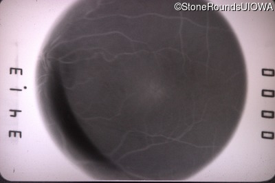
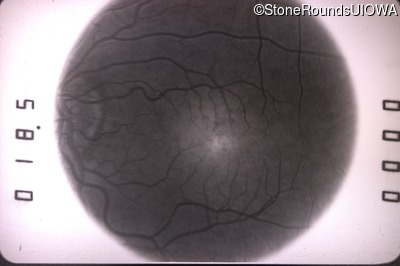
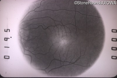
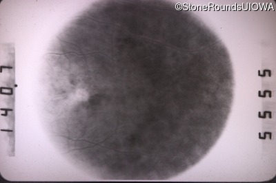
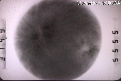
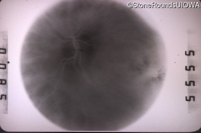
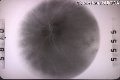
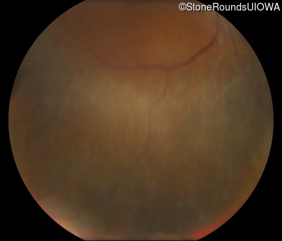
Visit at age: 28 years
Fundus Photography - Right - 20/20
Exemplar
Fundus Photography - Left - 20/50
Exemplar
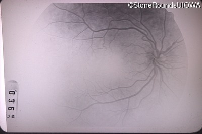
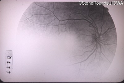
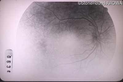
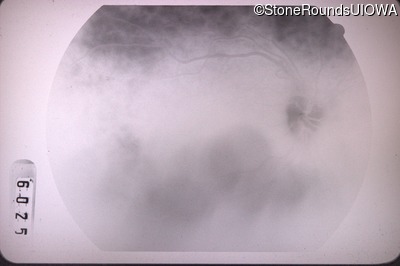
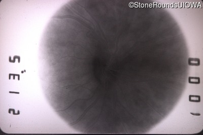
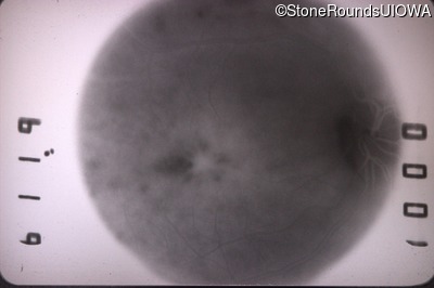
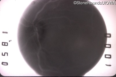
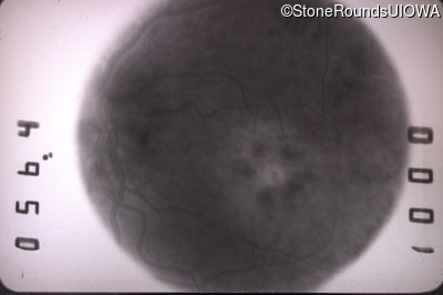
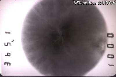
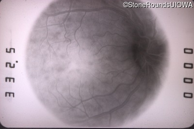
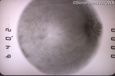
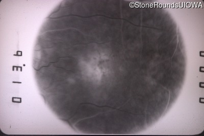
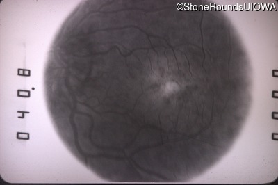
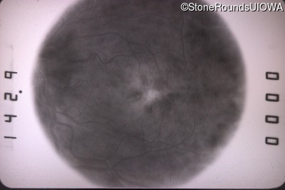
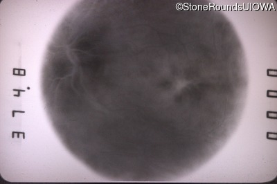
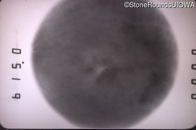
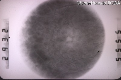
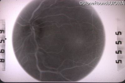
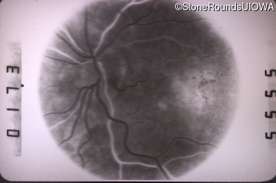
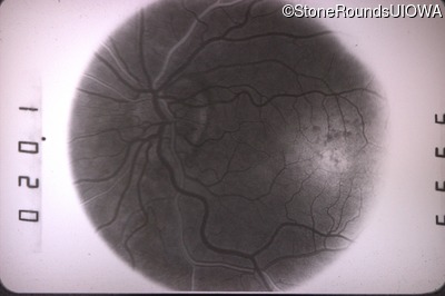
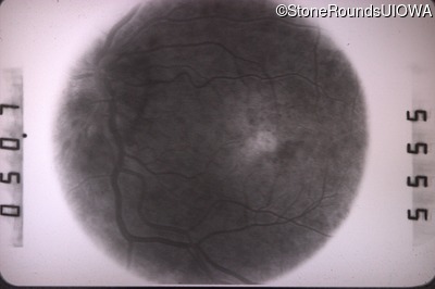
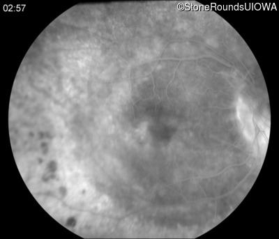
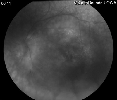
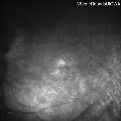
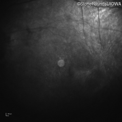
Fluorescein Angiography - Right - 20/20
Exemplar
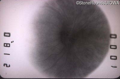
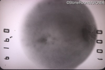
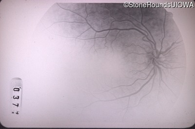
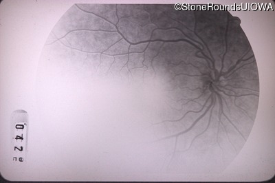
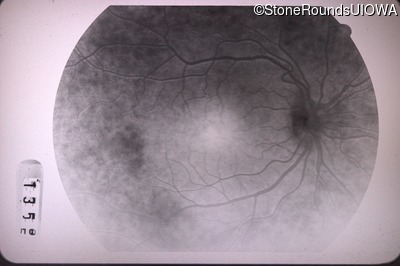
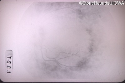
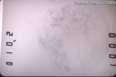
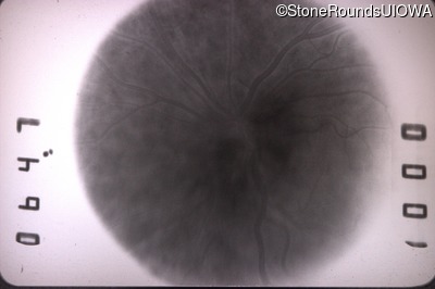
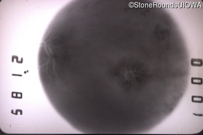
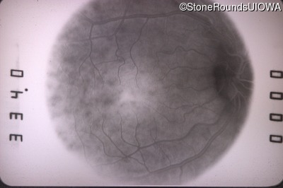
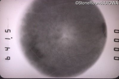
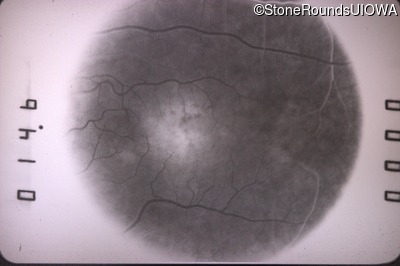
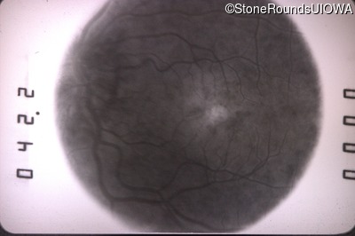
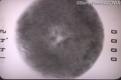
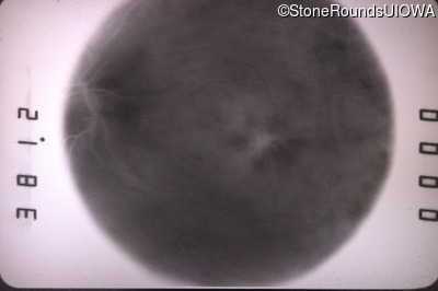
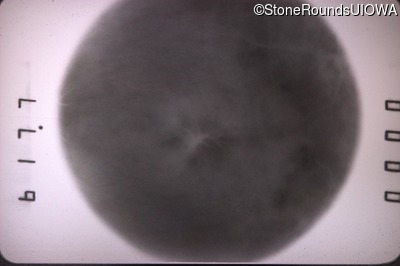
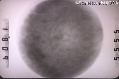
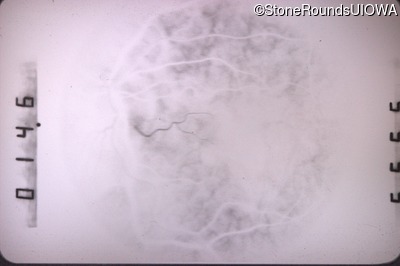
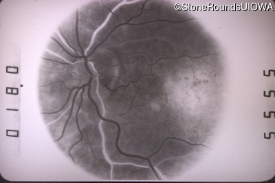
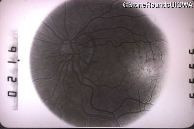
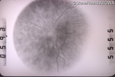
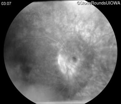
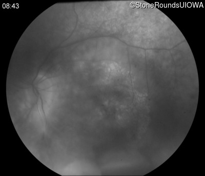
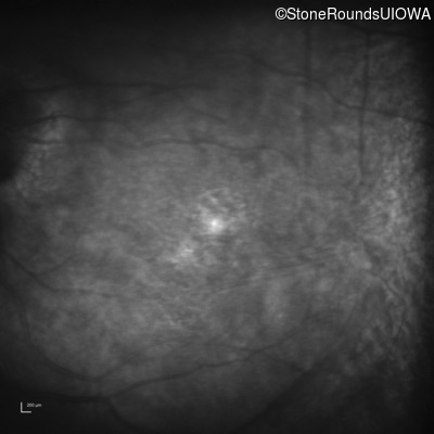
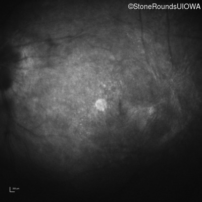
Fluorescein Angiography - Left - 20/50
Exemplar
Visit at age: 29 years
Fundus Photography - Right - 20/25 +2
Exemplar
Fundus Photography - Left - 20/40 -1
Exemplar
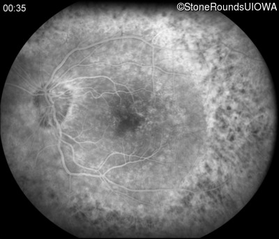
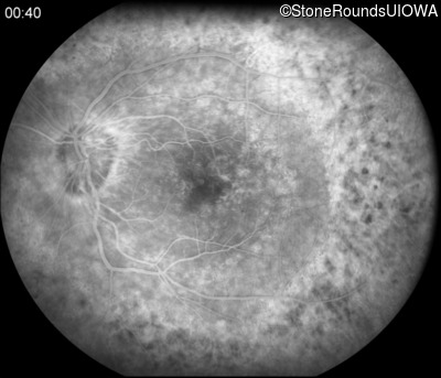
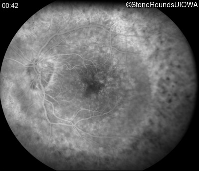
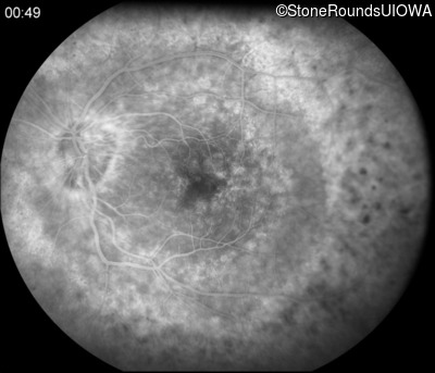
Fluorescein Angiography - Right - 20/25 +2
Exemplar
Fluorescein Angiography - Left - 20/40 -1
Exemplar
Visit at age: 29 years (Visit 2)
Fundus Photography - Right - 20/25
Exemplar
Fundus Photography - Left - 20/50
Exemplar
Fluorescein Angiography - Right - 20/25
Exemplar
Fluorescein Angiography - Left - 20/50
Exemplar

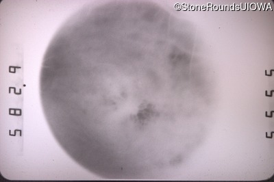
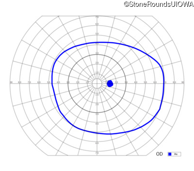
Visit at age: 33 years
Goldmann Visual Field - Right - 20/30 +1
Exemplar
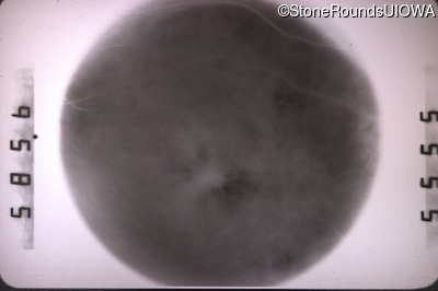
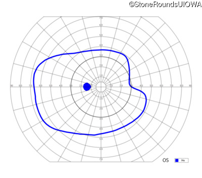
Goldmann Visual Field - Left - 20/70
Exemplar
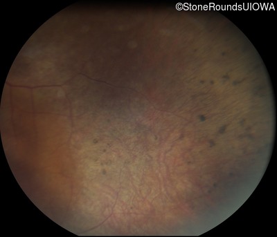
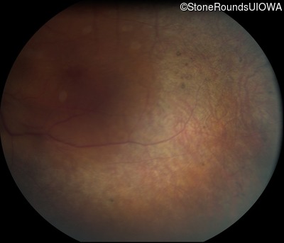
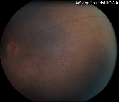
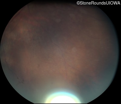
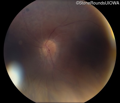
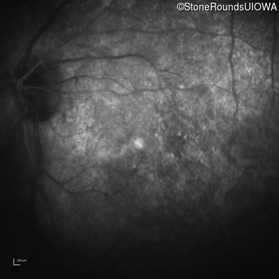
Visit at age: 42 years
Fundus Photography - Right - 20/40 -2
Exemplar
Fundus Photography - Left - 20/400
Exemplar

Fundus Montage - Right - 20/40 -2
Exemplar
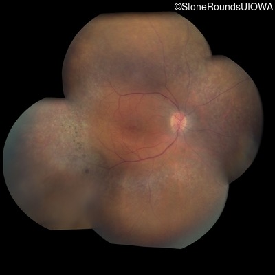
Fundus Montage - Left - 20/400
Exemplar
Fluorescein Angiography - Right - 20/40 -2
Exemplar
Fluorescein Angiography - Left - 20/400
Exemplar


Visit at age: 42 years (Visit 2)
Fundus Photography - Right - 20/40 -1
Exemplar
Fundus Photography - Left - 20/300
Exemplar
Visit at age: 43 years
Fundus Photography - Right - 20/40
Exemplar
Fundus Photography - Left - 20/300
Exemplar
Visit at age: 43 years (Visit 2)
Fundus Photography - Right - 20/40
Exemplar
Fundus Photography - Left - 20/250
Exemplar
Visit at age: 43 years (Visit 3)
Fundus Photography - Right - 20/30
Exemplar
Visit at age: 44 years
Fundus Photography - Right - 20/40 -1
Exemplar
Fundus Photography - Left - 20/200
Exemplar
Visit at age: 44 years (Visit 2)
Fundus Photography - Right - 20/60
Exemplar
Fundus Photography - Left - 20/200
Exemplar
Visit at age: 45 years
Fundus Photography - Right - 20/50 +2
Exemplar
Fundus Photography - Left - 20/160
Exemplar
Fluorescein Angiography - Right - 20/50 +2
Exemplar


Fluorescein Angiography - Left - 20/160
Exemplar
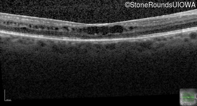
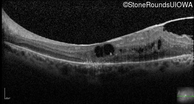
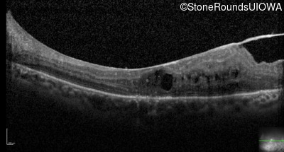
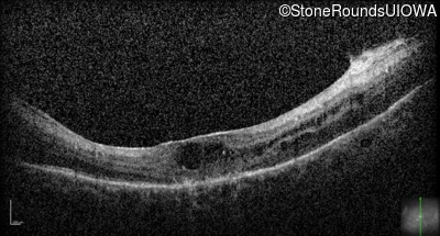
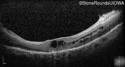
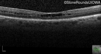
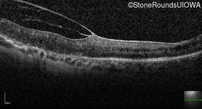
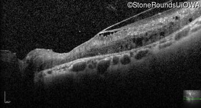
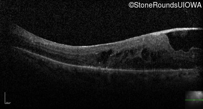
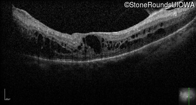
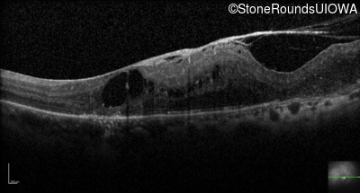
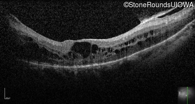
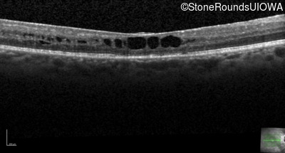
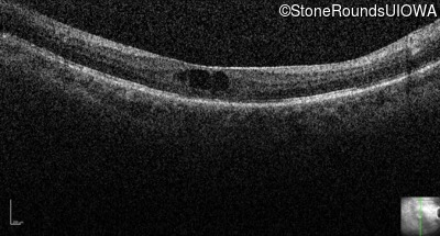
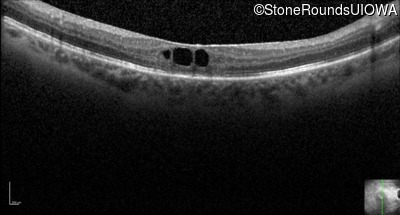
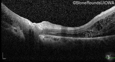
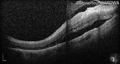
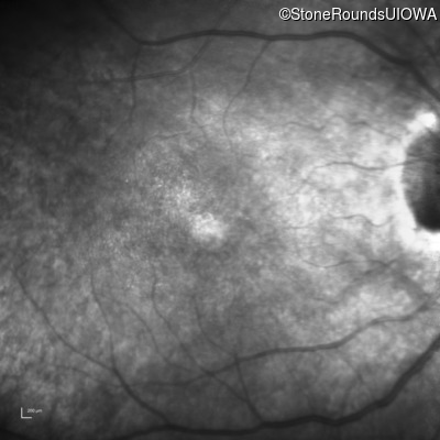
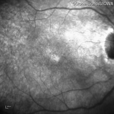
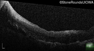
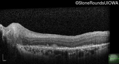
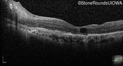
Visit at age: 45 years (Visit 2)
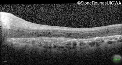
Optical Coherence Tomography - Right - 20/50 +2
Exemplar / OCT Stack
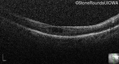
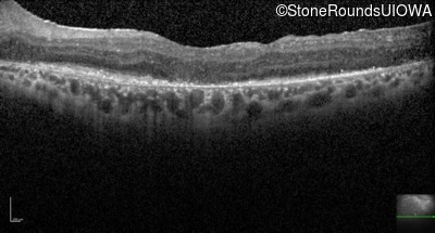
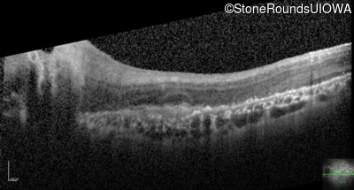
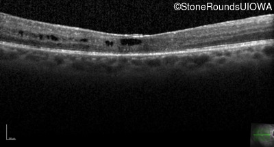
Optical Coherence Tomography - Left - 20/160
Exemplar / OCT Stack
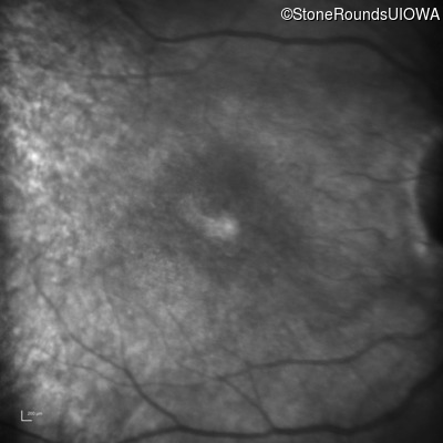
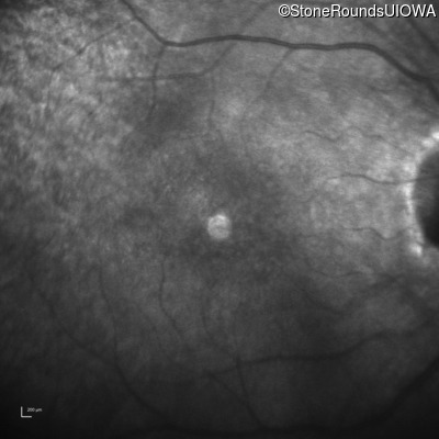
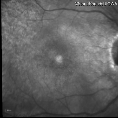
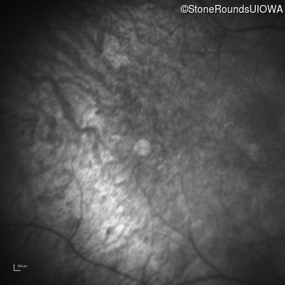
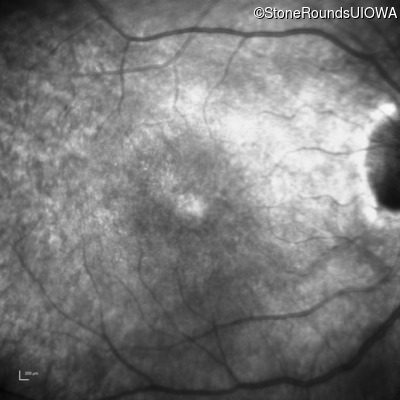
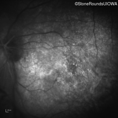
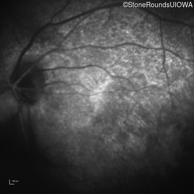
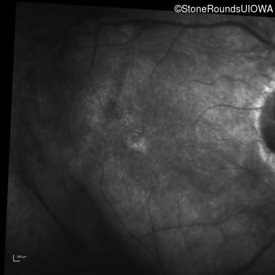
Infrared Fundus Photograph - Right - 20/50 +2
Exemplar
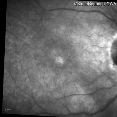
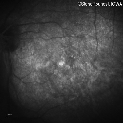
Infrared Fundus Photograph - Left - 20/160
Exemplar
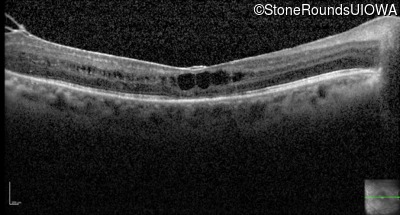
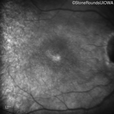
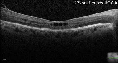
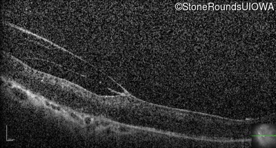
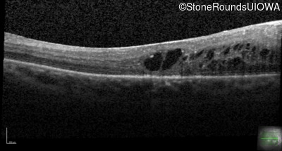
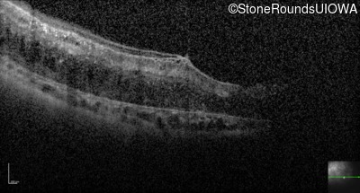
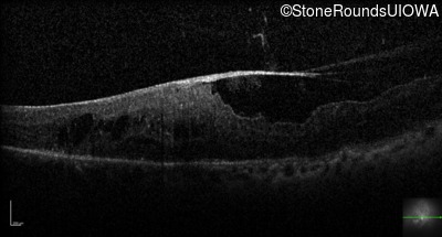
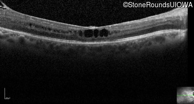
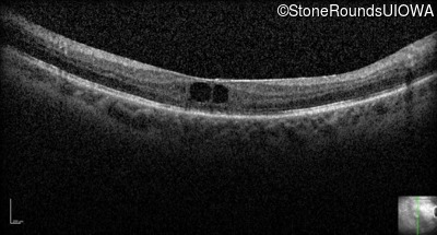
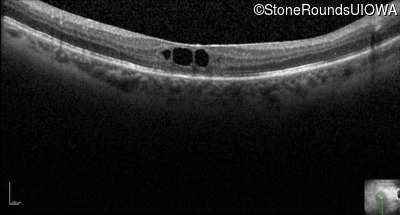
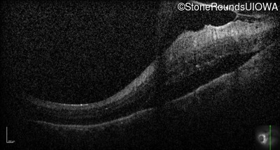
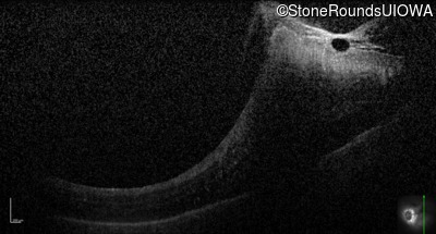
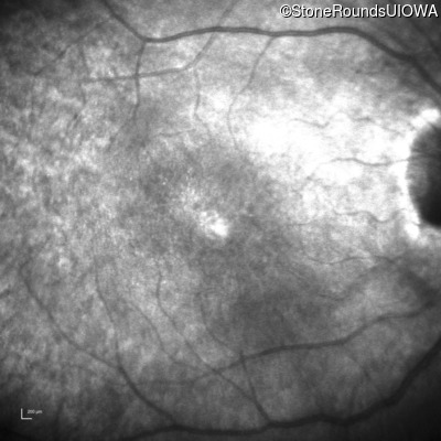
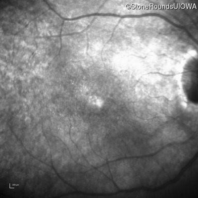
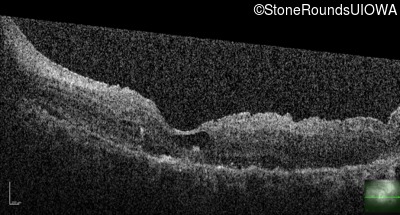
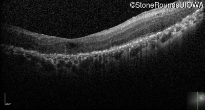
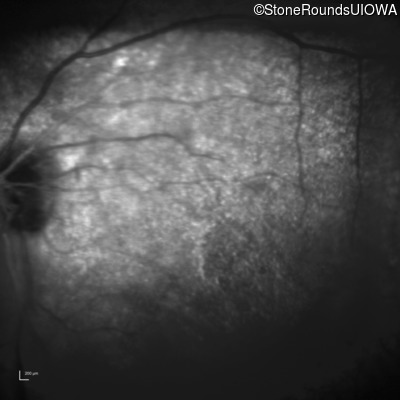
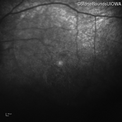
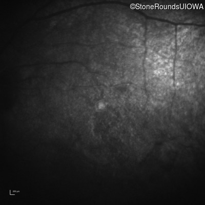
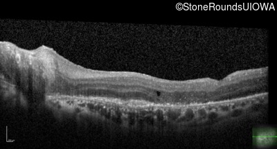
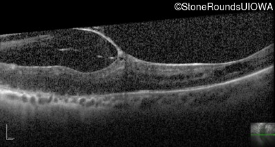
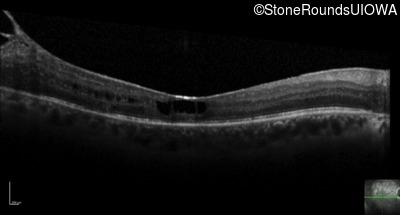
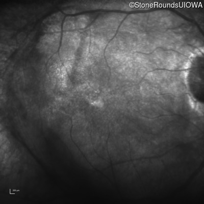
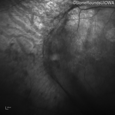
Visit at age: 46 years
Optical Coherence Tomography - Right - 20/50 +2
Exemplar / OCT Stack
Optical Coherence Tomography - Left - 20/160
Exemplar / OCT Stack
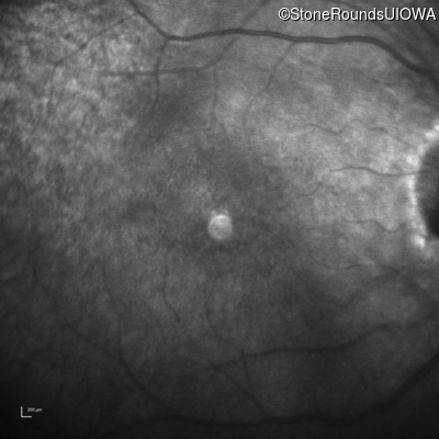
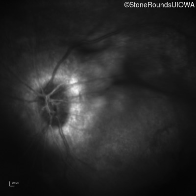
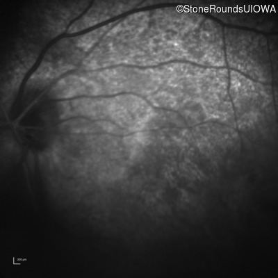
Infrared Fundus Photograph - Right - 20/50 +2
Exemplar
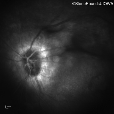
Infrared Fundus Photograph - Left - 20/160
Exemplar
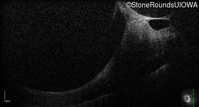
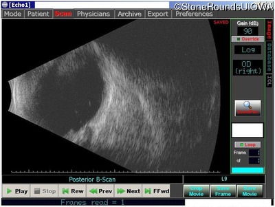
Visit at age: 46 years (Visit 2)
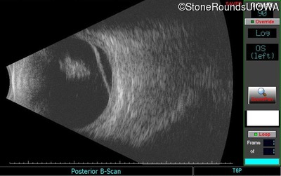
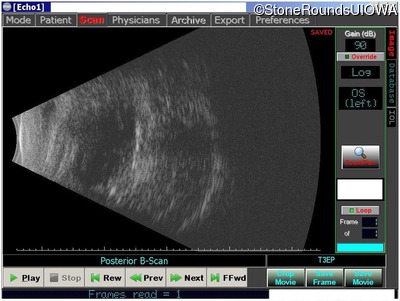
B-Scan Ultrasonography - Left - unknown
Exemplar
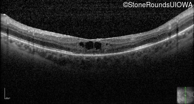
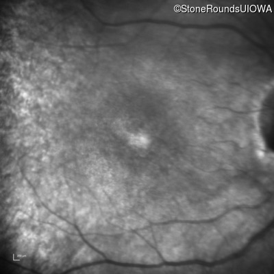
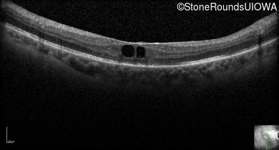
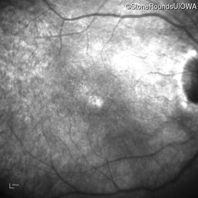
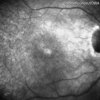
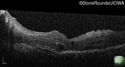
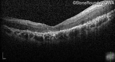
Visit at age: 46 years (Visit 3)
Fundus Photography - Right - 20/50 -3
Exemplar
Fundus Photography - Left - Count Fingers 6"
Exemplar
Optical Coherence Tomography - Right - 20/50 -3
Exemplar / OCT Stack

Optical Coherence Tomography - Left - Count Fingers 6"
Exemplar / OCT Stack
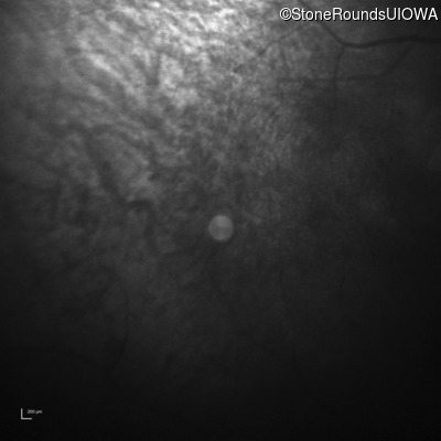
Infrared Fundus Photograph - Right - 20/50 -3
Exemplar

Infrared Fundus Photograph - Left - Count Fingers 6"
Exemplar
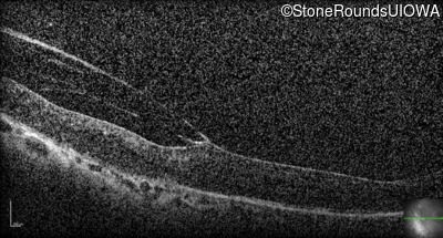
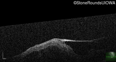
Visit at age: 46 years (Visit 4)
Optical Coherence Tomography - Left - Hand Motion
Exemplar / OCT Stack
OCT Stack
OCT Stack
OCT Stack
Infrared Fundus Photograph - Left - Hand Motion
Exemplar
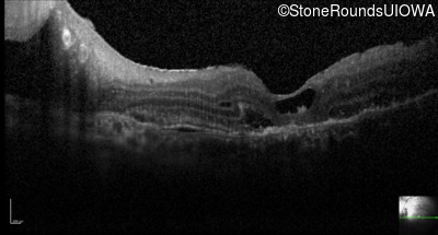
Visit at age: 46 years (Visit 5)
Fundus Photography - Left - Hand Motion
Exemplar
Optical Coherence Tomography - Left - Hand Motion
Exemplar / OCT Stack
Infrared Fundus Photograph - Left - Hand Motion
Exemplar
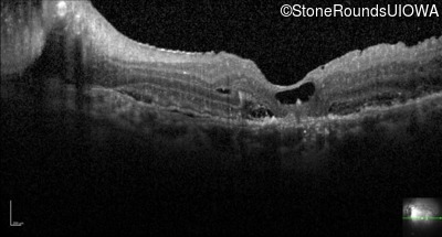
Visit at age: 46 years (Visit 6)
Optical Coherence Tomography - Left - Count Fingers 3'
Exemplar / OCT Stack
Infrared Fundus Photograph - Left - Count Fingers 3'
Exemplar
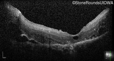
Visit at age: 46 years (Visit 7)
Optical Coherence Tomography - Left - Light Perception
Exemplar / OCT Stack
OCT Stack
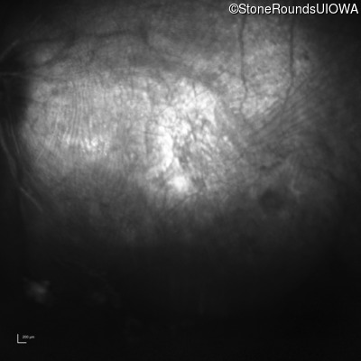
Infrared Fundus Photograph - Left - Light Perception
Exemplar
Visit at age: 49 years
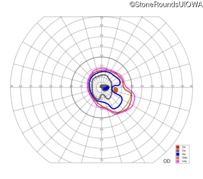
Goldmann Visual Field - Right - 20/70
Exemplar
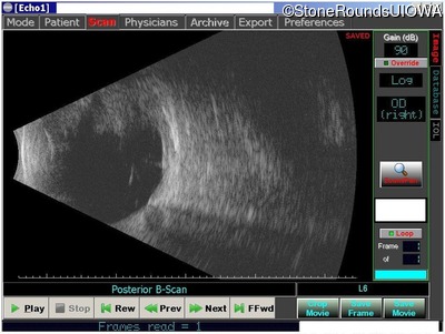
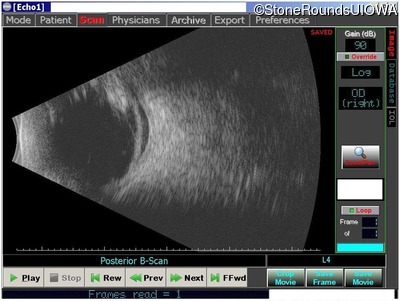
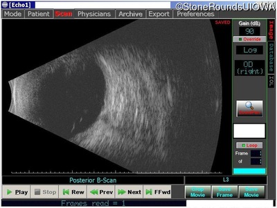
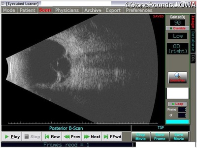
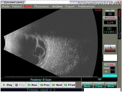
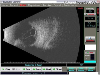
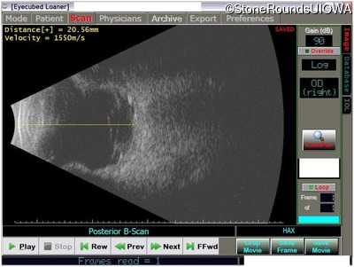
B-Scan Ultrasonography - Right - 20/70
Exemplar

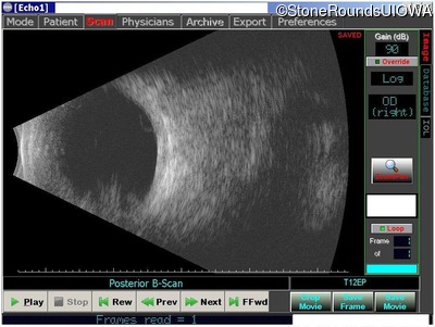
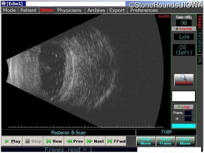
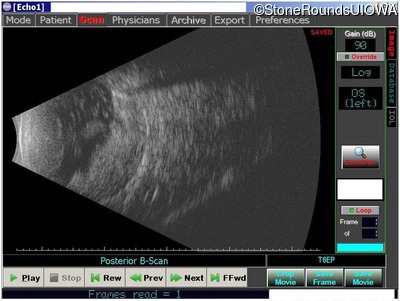
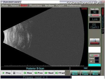
B-Scan Ultrasonography - Left - No Light Perception
Exemplar
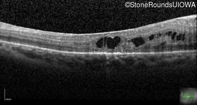
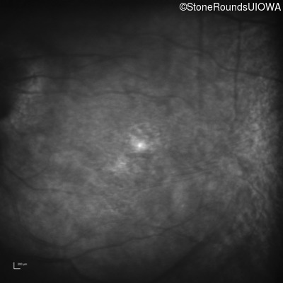
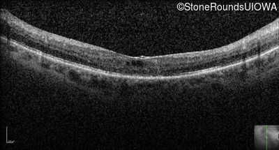
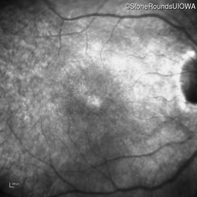
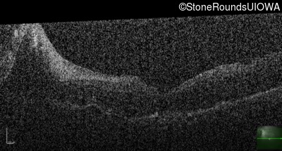
Visit at age: 50 years
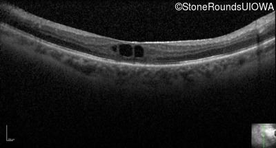
Optical Coherence Tomography - Right - 20/50 -2
Exemplar / OCT Stack
OCT Stack
OCT Stack
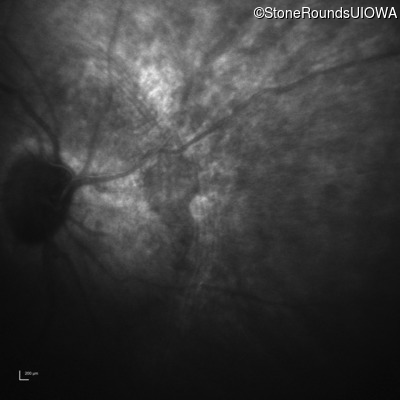
Infrared Fundus Photograph - Right - 20/50 -2
Exemplar
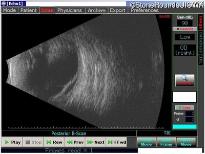
Visit at age: 51 years
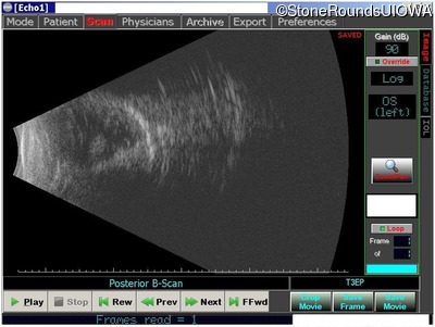
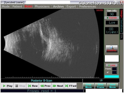
B-Scan Ultrasonography - Right - 20/400
Exemplar
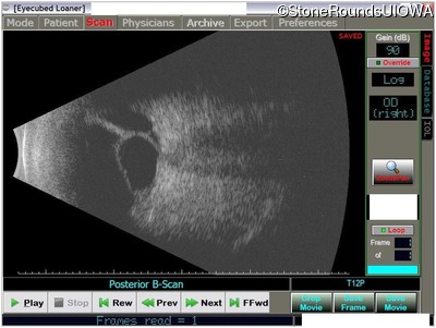
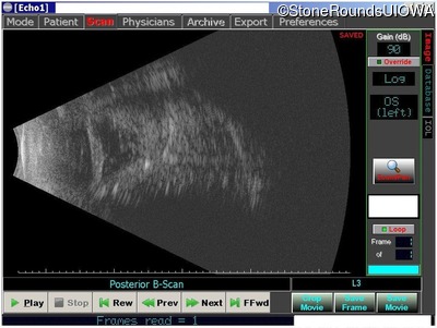
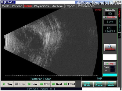
B-Scan Ultrasonography - Left - No Light Perception
Exemplar
Case Level Images
Diagnosis & molecular findings
| Disease | Gene | Allele 1 variant(s) | Allele 2 variant(s) | Inheritance mode |
|---|---|---|---|---|
| AD Neovascular Inflammatory Vitreoretinopathy | CAPN5 | Arg243Leu CGC>CTC | AD |
Gene:
Allele 1:
Arg243Leu CGC>CTC
Allele 2:
Inheritance:
AD