Case
SR61
Student Mode
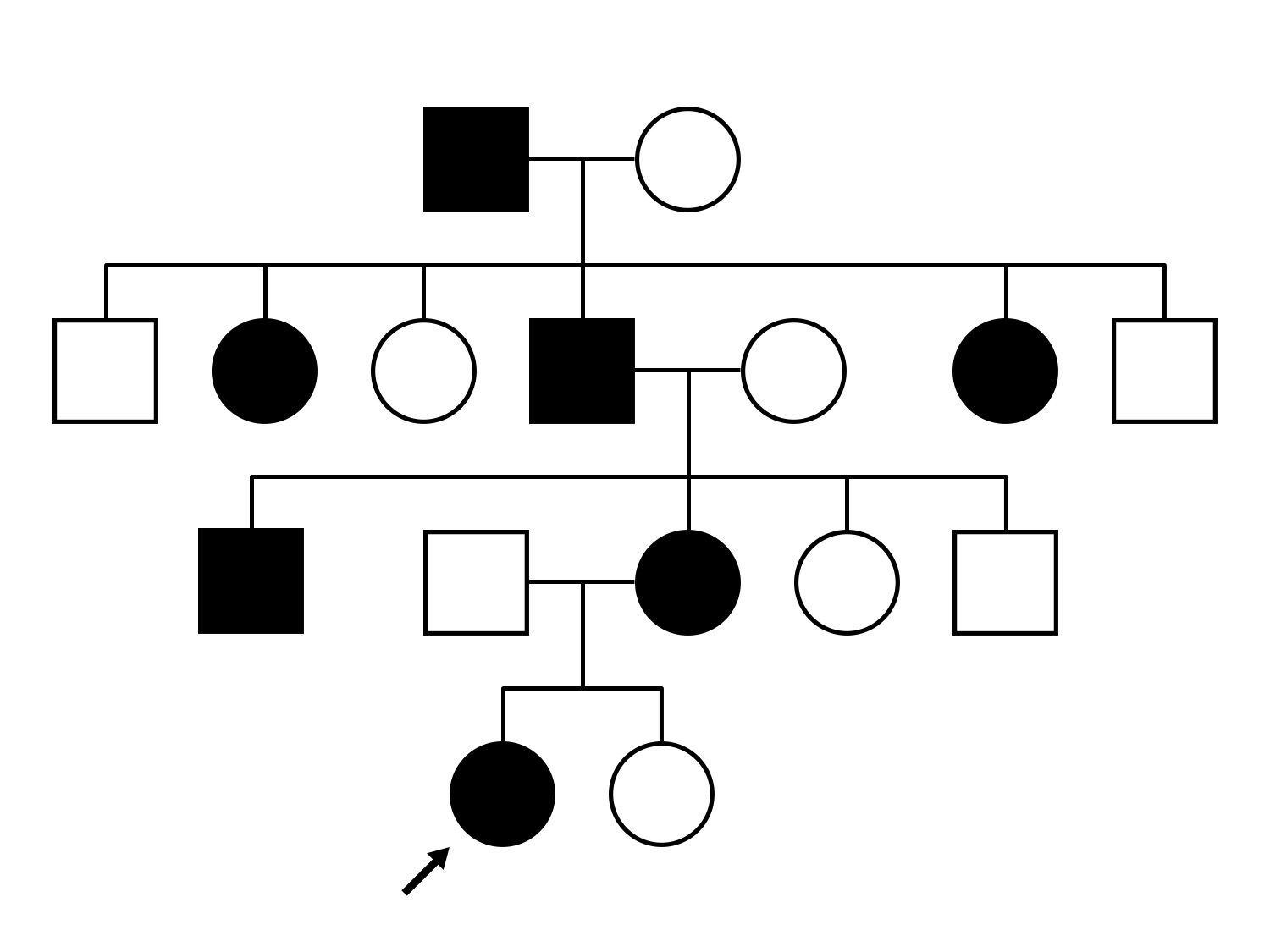
AD Neovascular Inflammatory Vitreoretinopathy (IIIE3)
Female
Female
Hidden
SR61
Student Mode
AD Neovascular Inflammatory Vitreoretinopathy (IIIE3)
Female
Female
Please Login or Register to download images or create a PowerPoint slideshow.
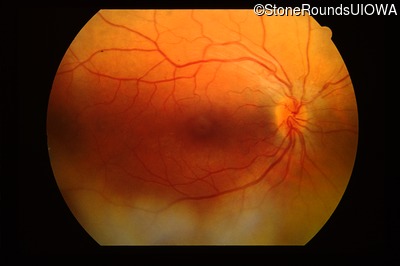
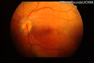
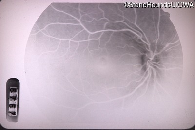
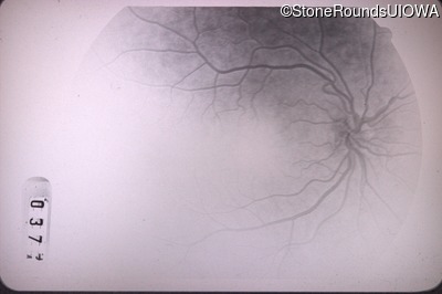
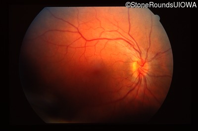
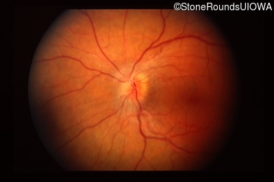
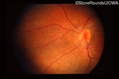
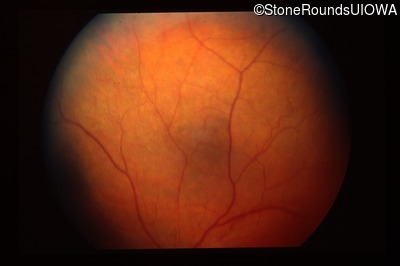
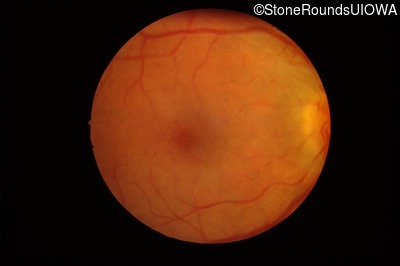
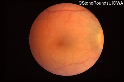
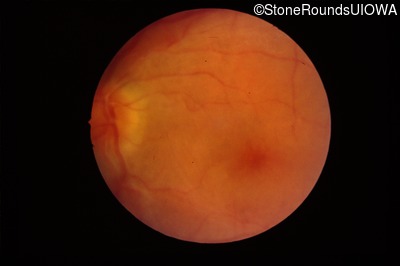
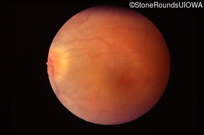
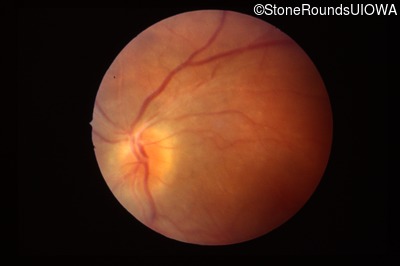
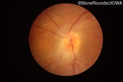
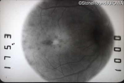
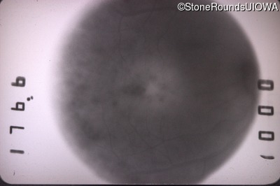
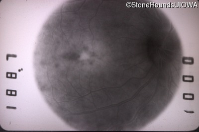
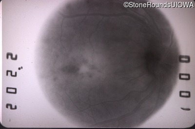
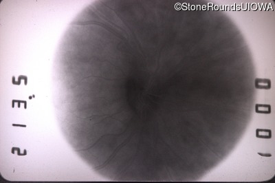
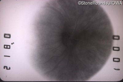
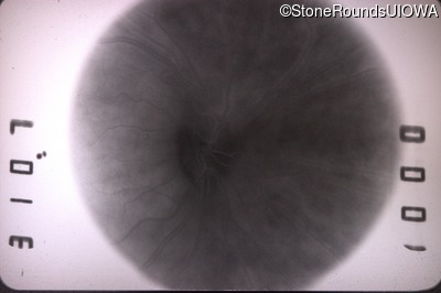
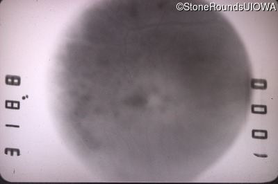
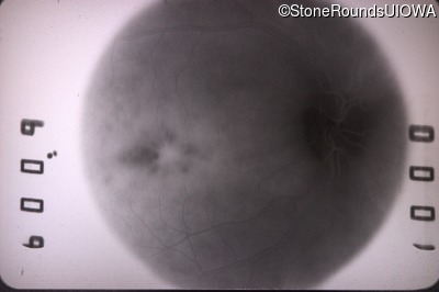
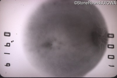
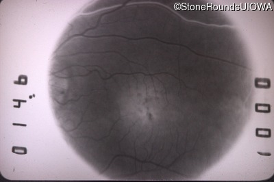
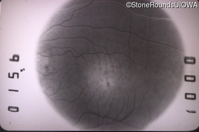
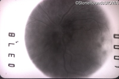
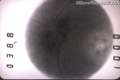
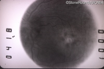
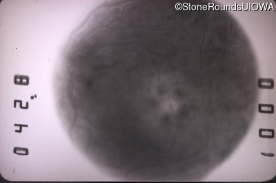
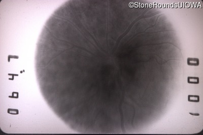
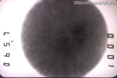
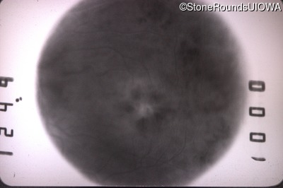
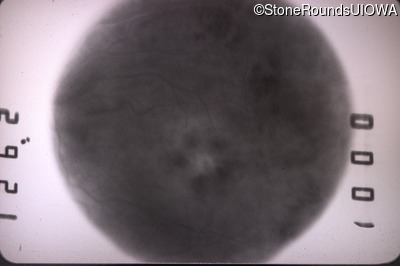
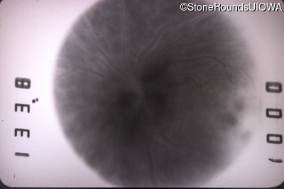
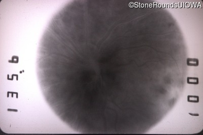
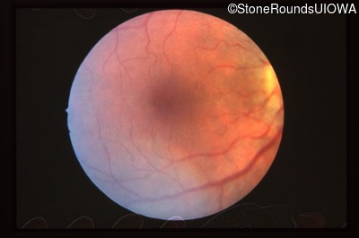
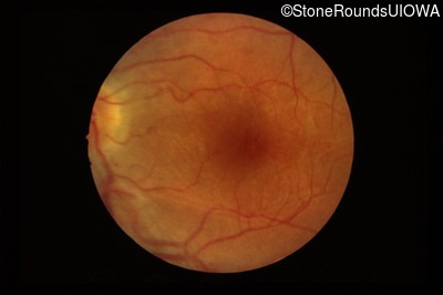
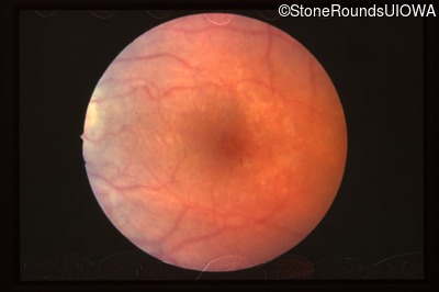
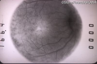
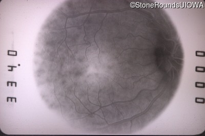
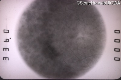
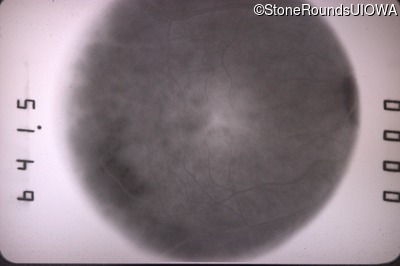
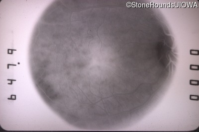
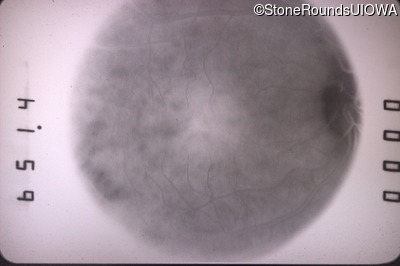
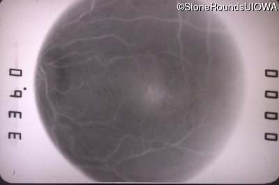
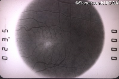
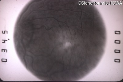
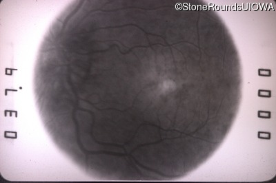
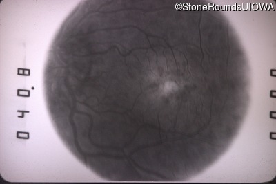
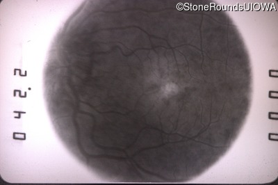
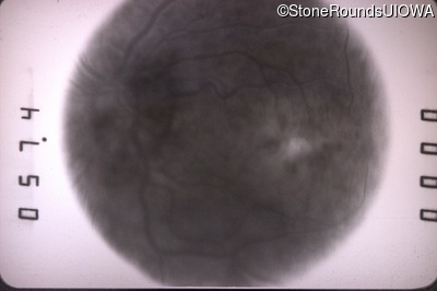
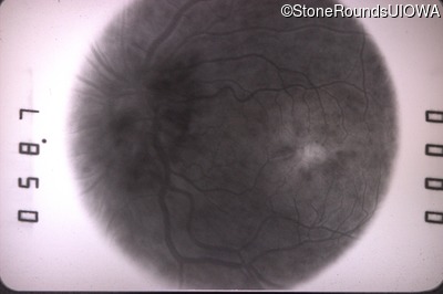
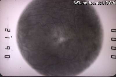
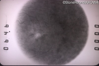
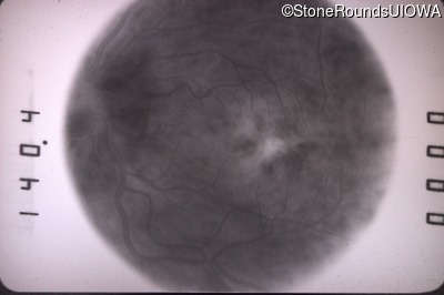
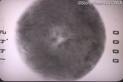
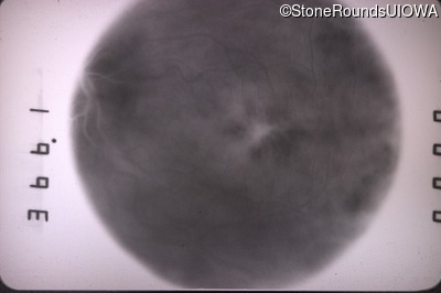
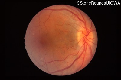
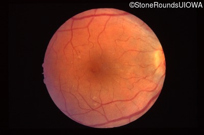
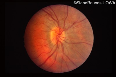
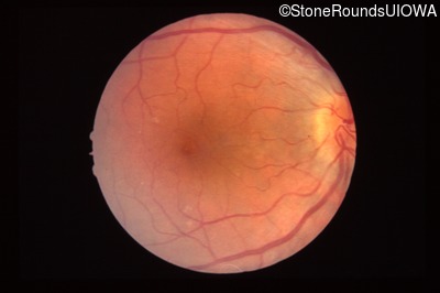
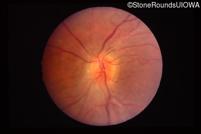
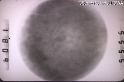
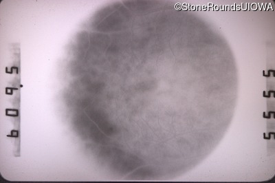
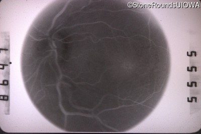
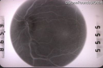
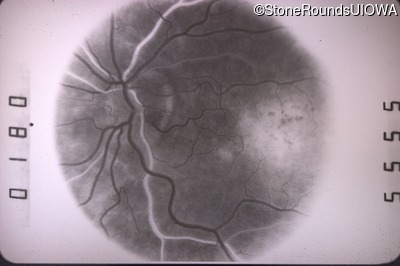
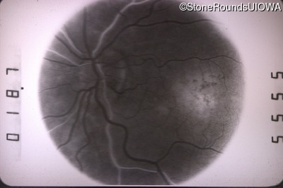
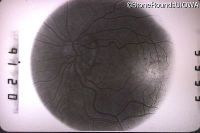
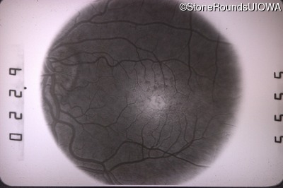
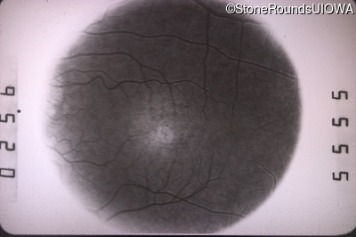
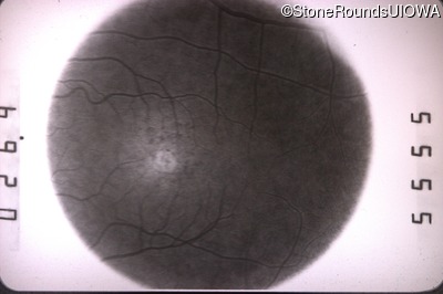
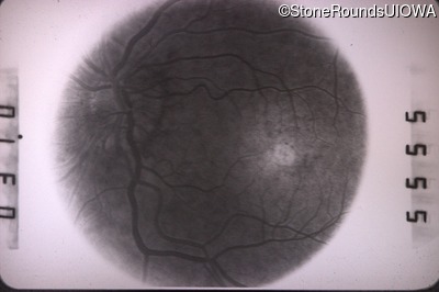
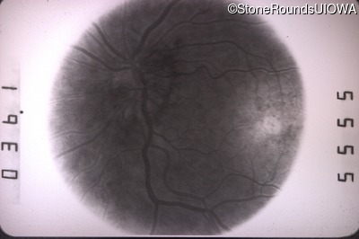
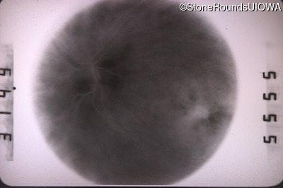
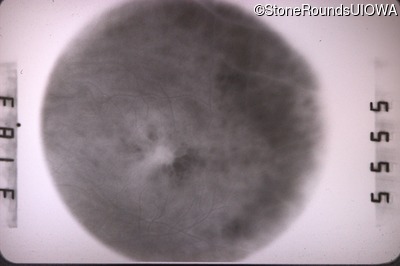
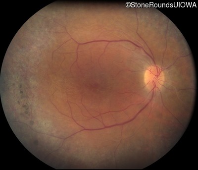
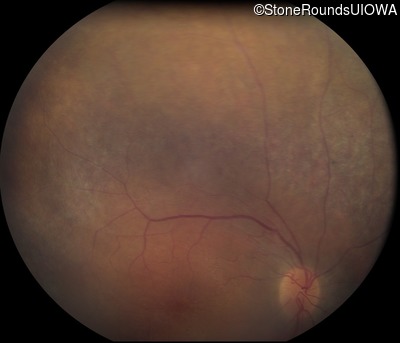
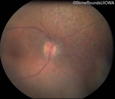
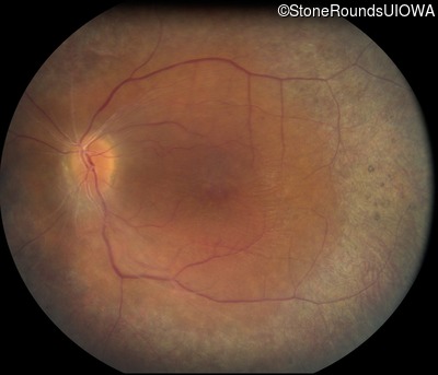
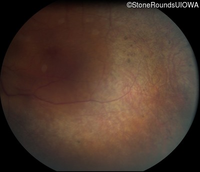
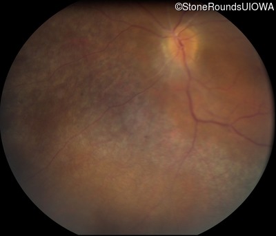
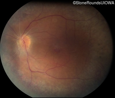
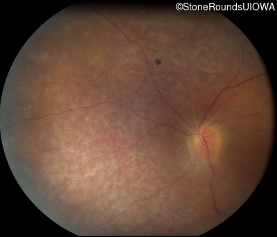
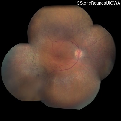
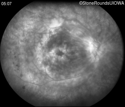
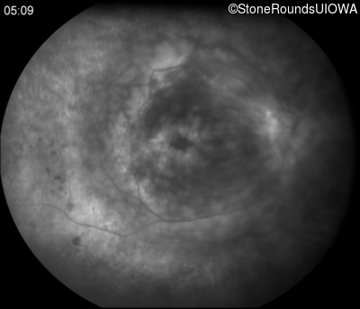
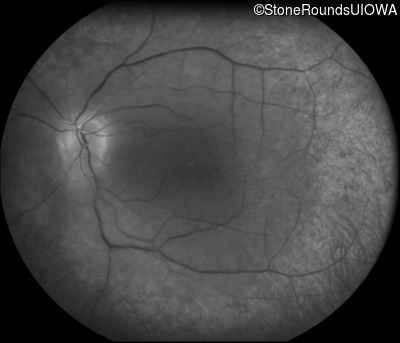
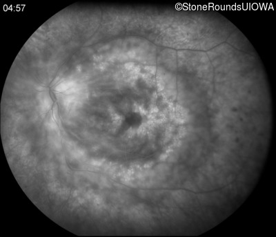
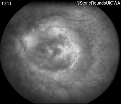
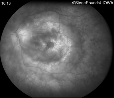
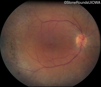
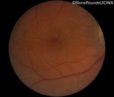
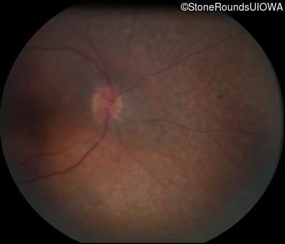
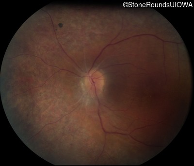
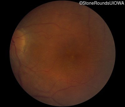
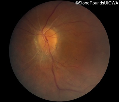
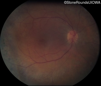
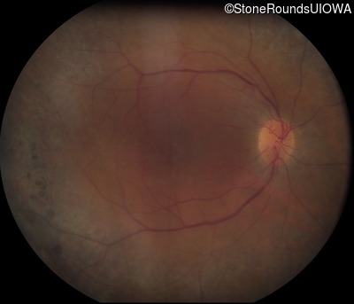
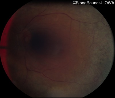
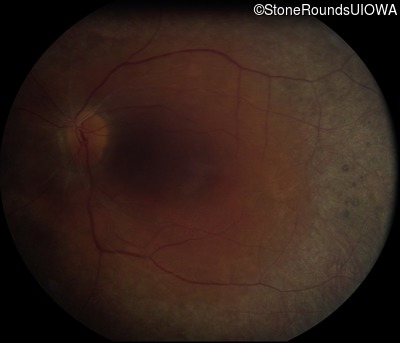
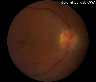
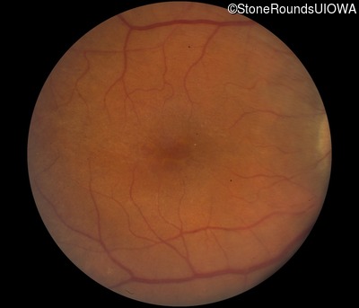
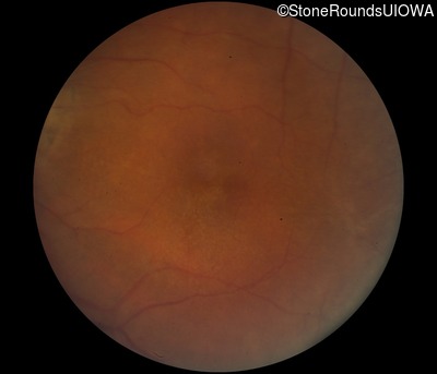
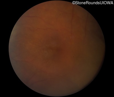
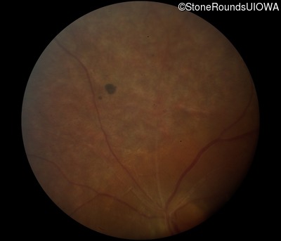
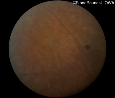
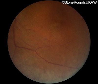
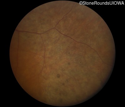
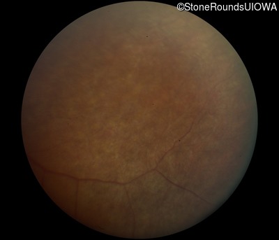
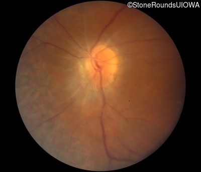
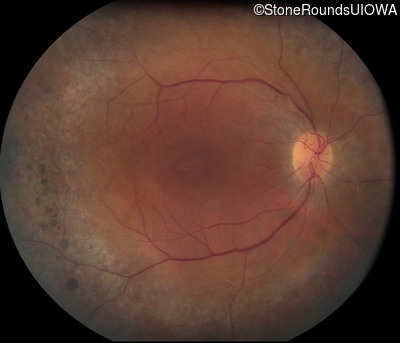
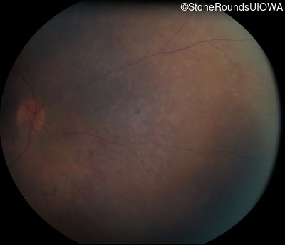
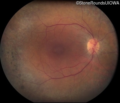
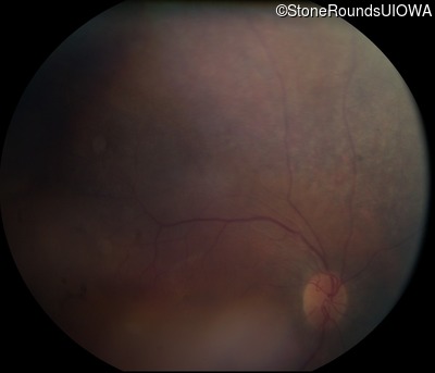
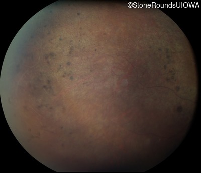
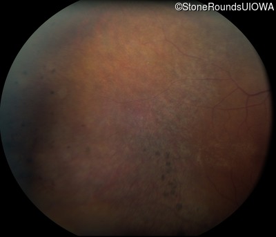
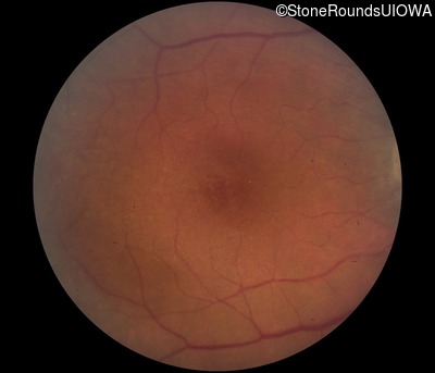
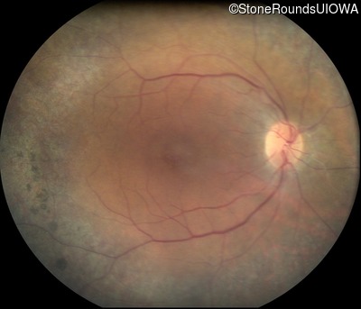
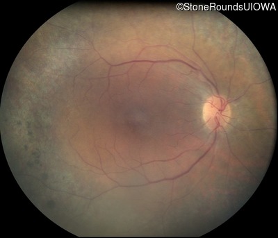
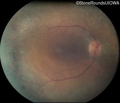
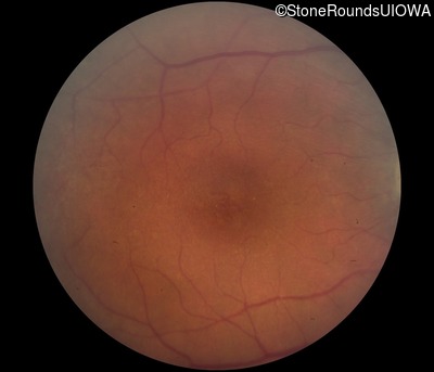
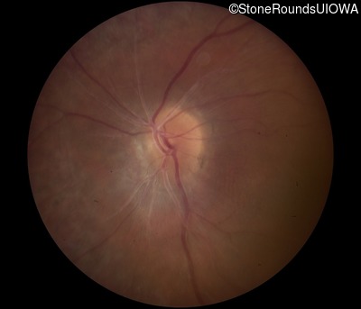
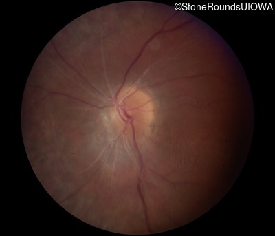
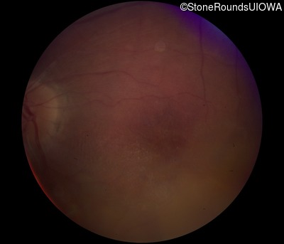
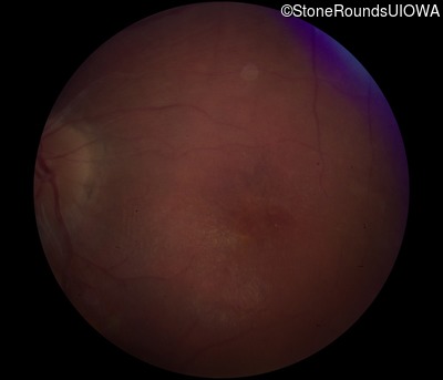
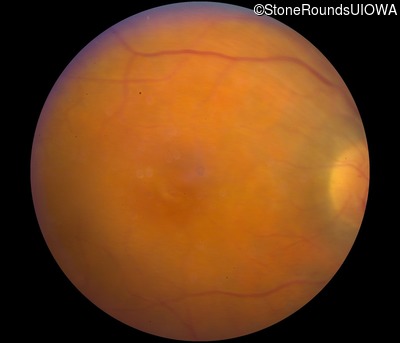
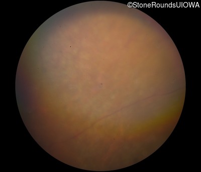
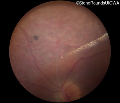
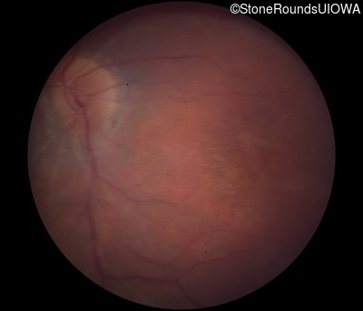
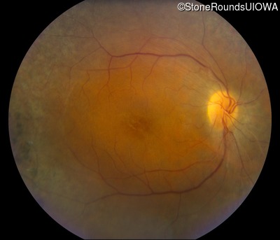
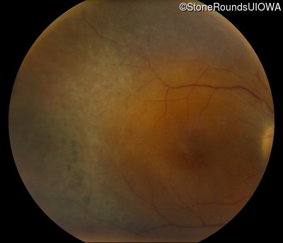
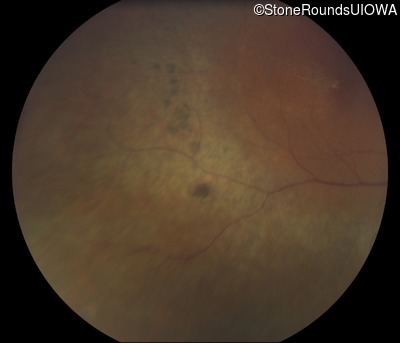
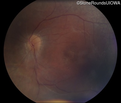
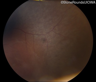
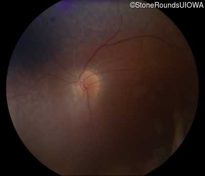
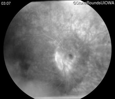
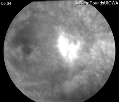
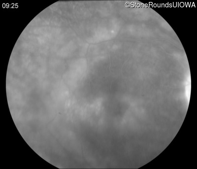
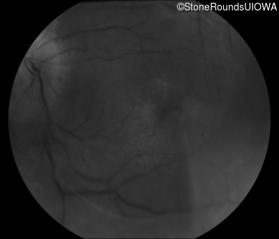
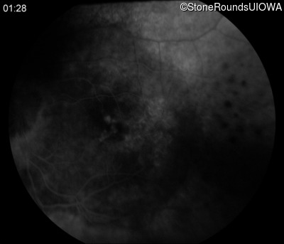
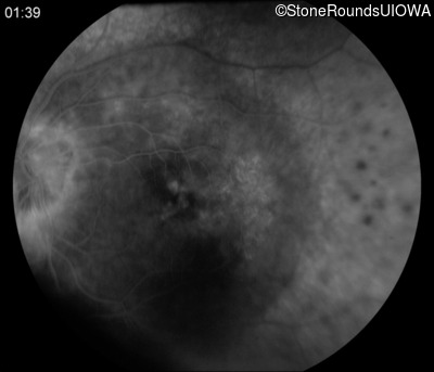
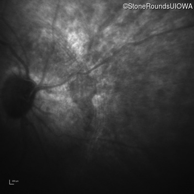
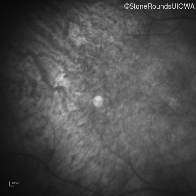
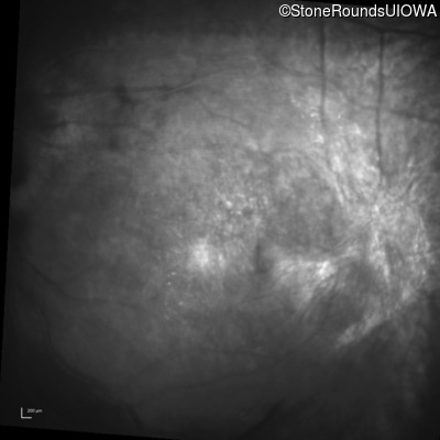
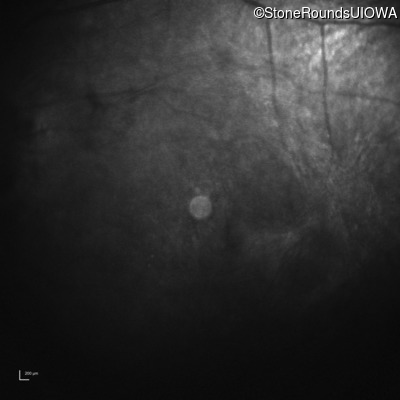
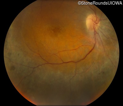
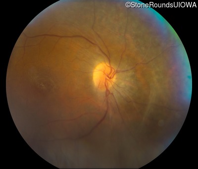
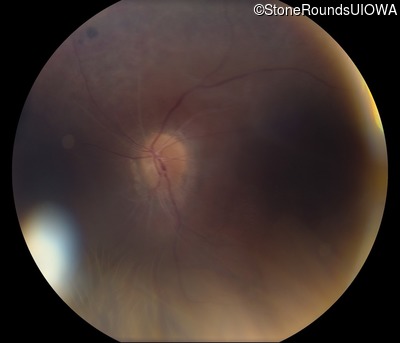
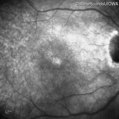
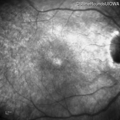
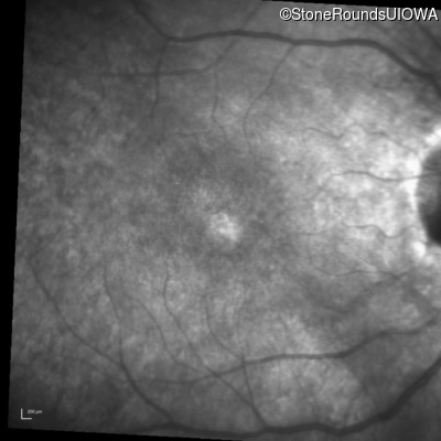
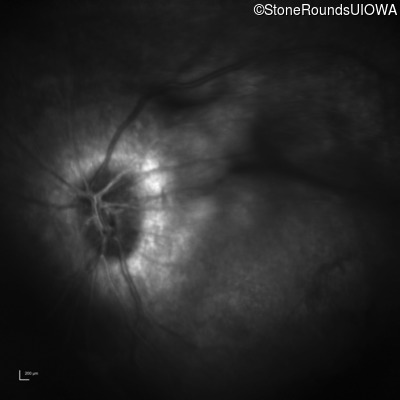
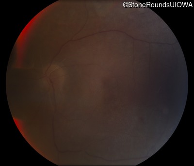
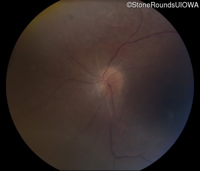
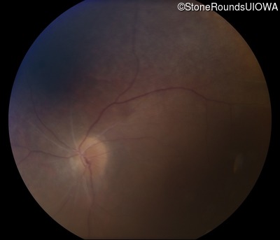
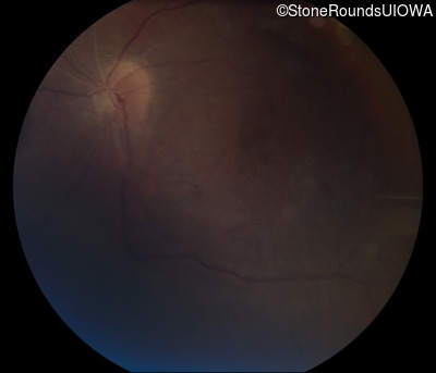
Visit at age: 23 years
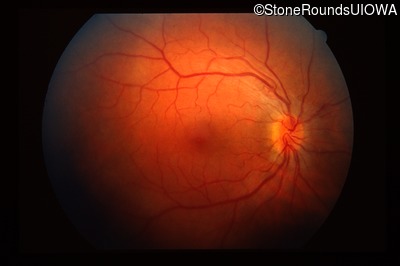
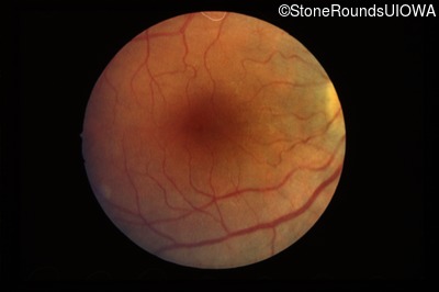
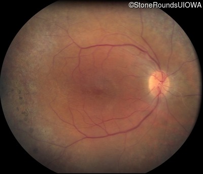
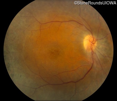
Fundus Photography - Right - 20/40
Exemplar
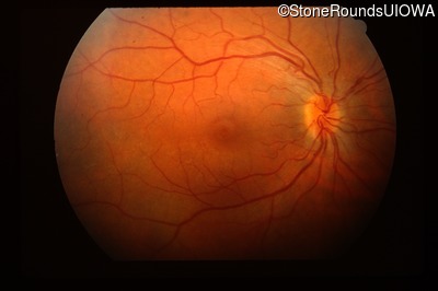
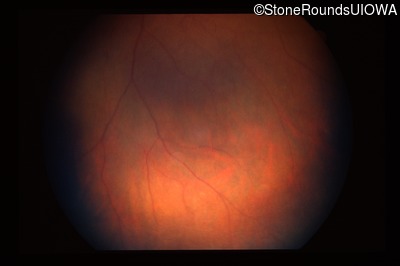
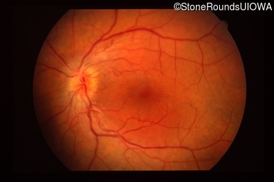
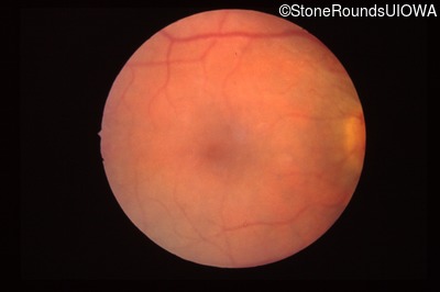
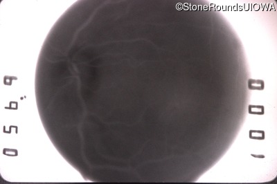
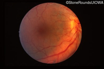
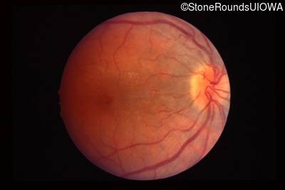
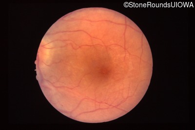
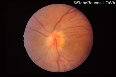
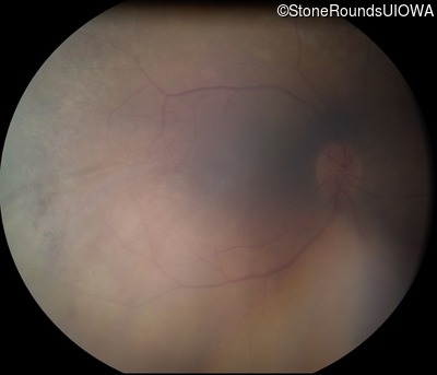
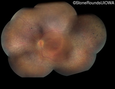
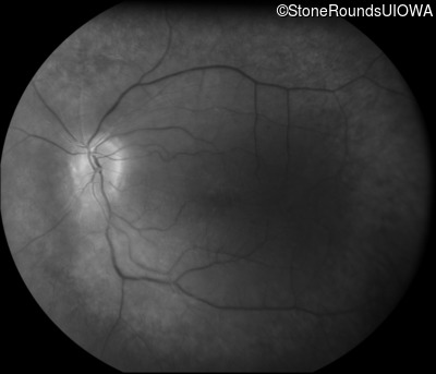
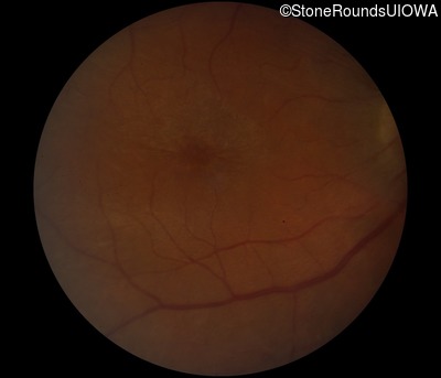
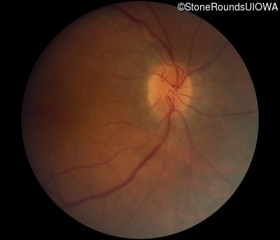
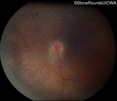
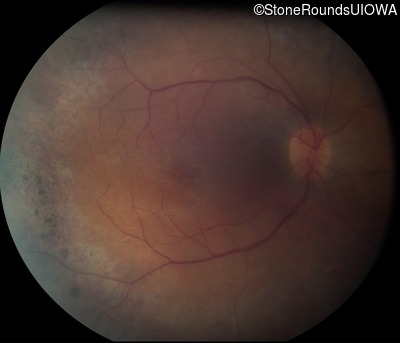
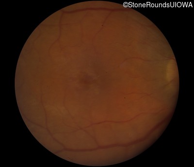
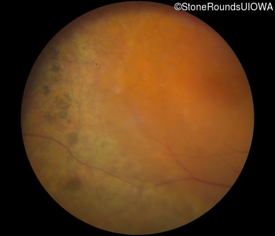
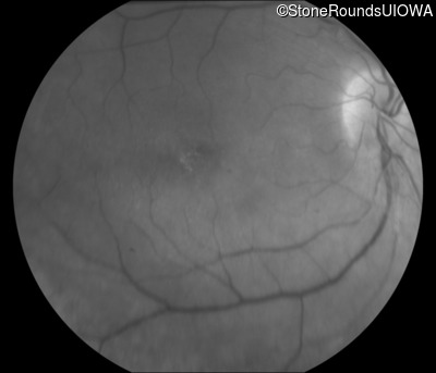
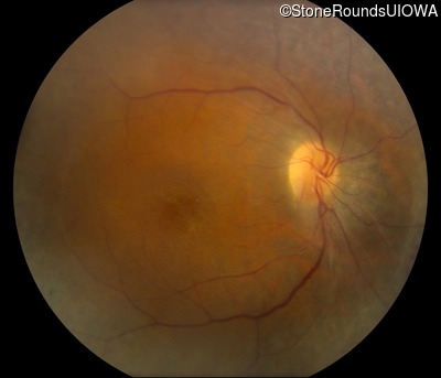
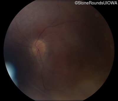
Fundus Photography - Left - 20/30
Exemplar
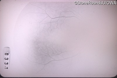
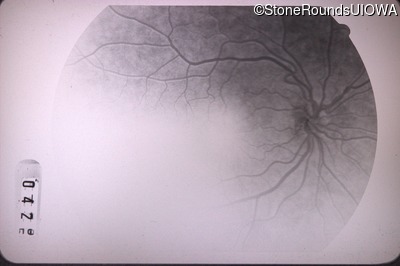
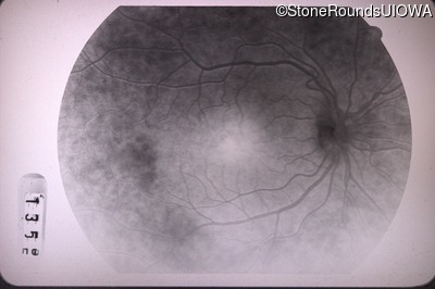
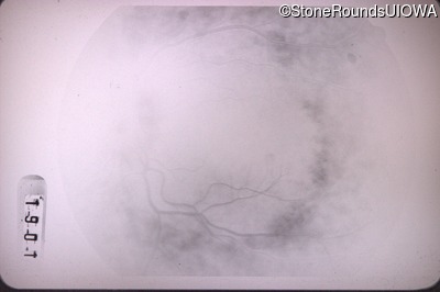
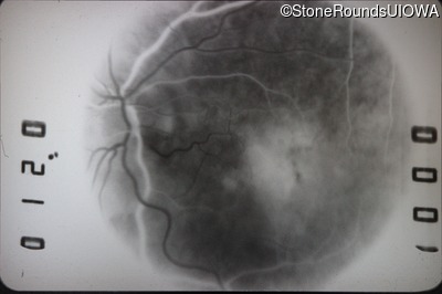
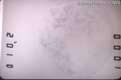
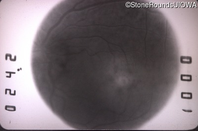
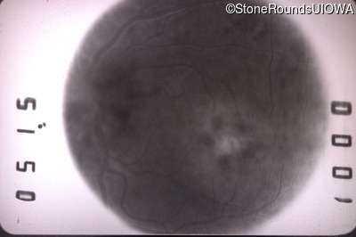
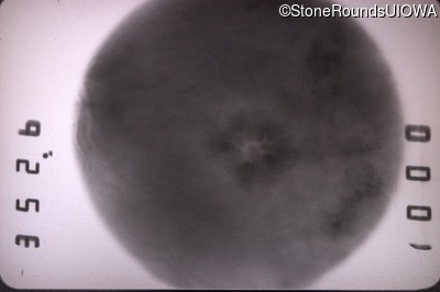
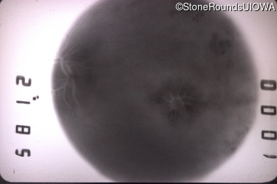
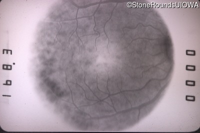
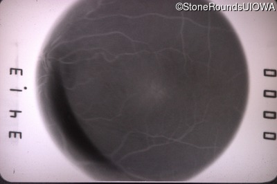
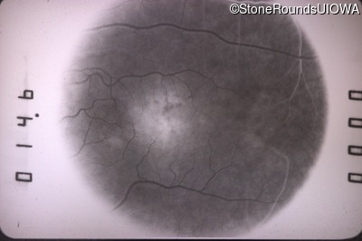
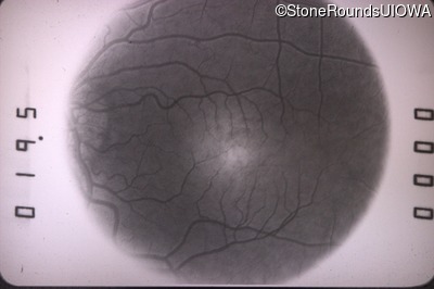
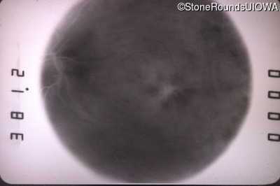
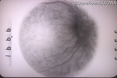
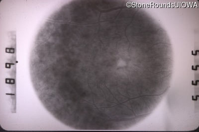
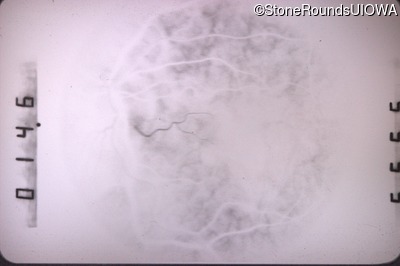
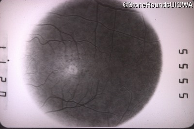
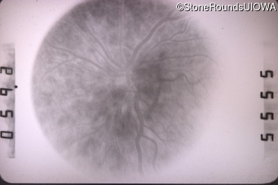
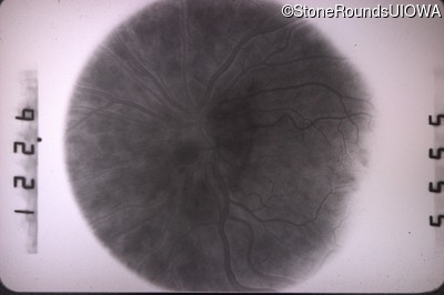
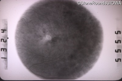
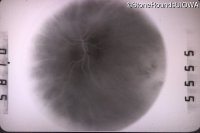
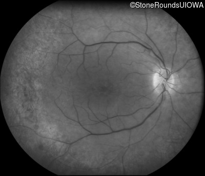
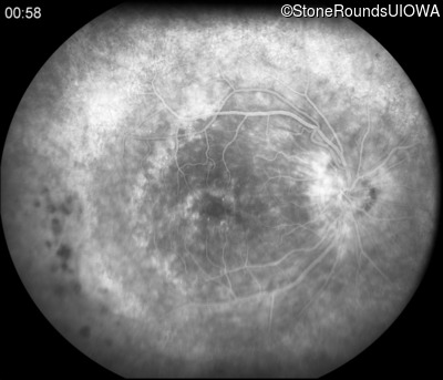
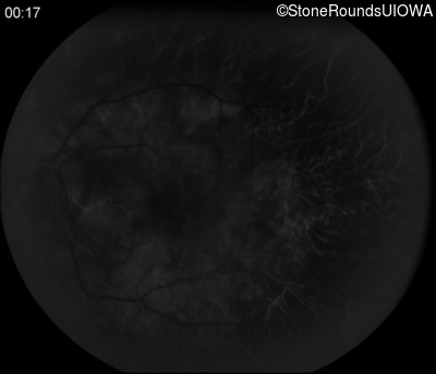
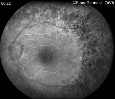
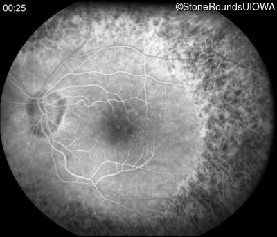
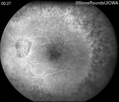
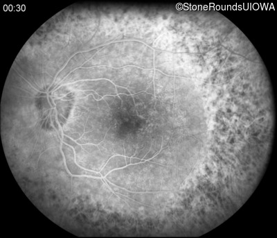
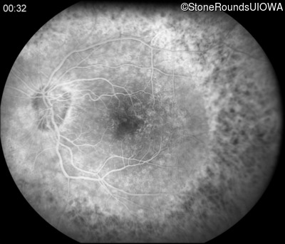
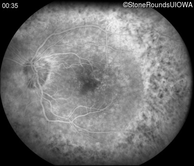
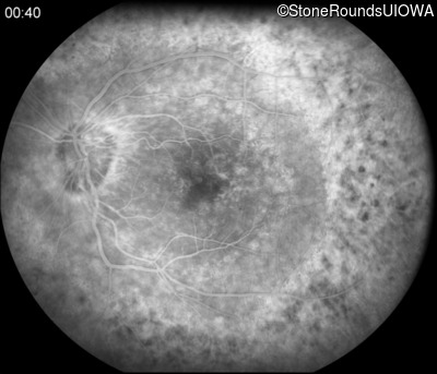
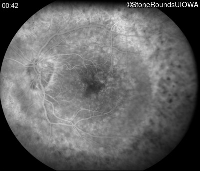
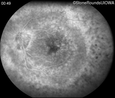
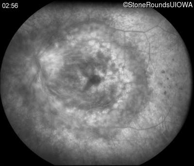
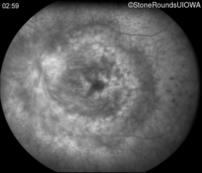
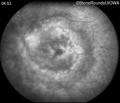
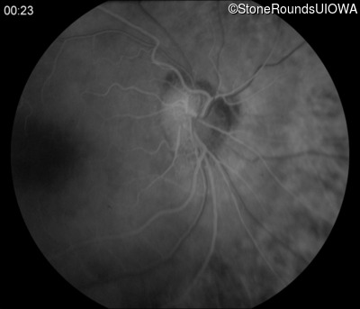
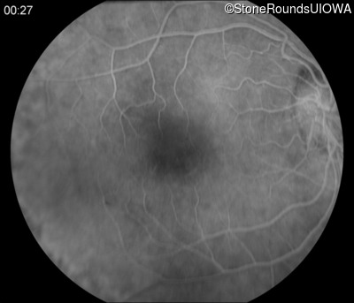
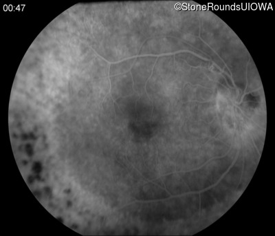
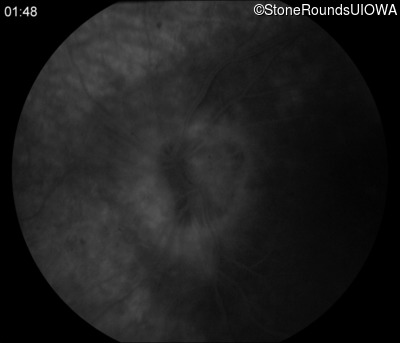
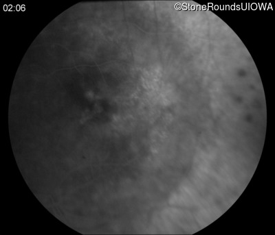
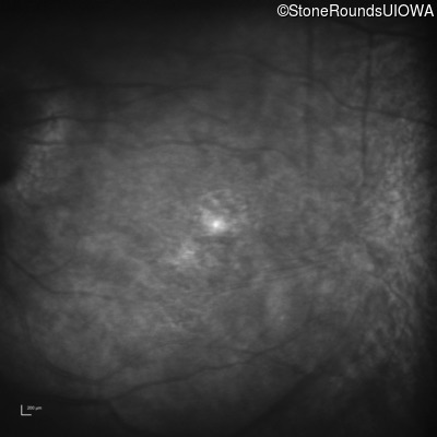
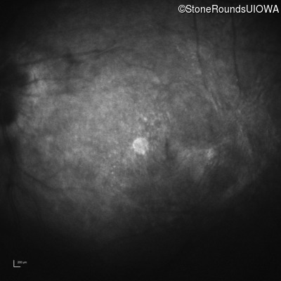
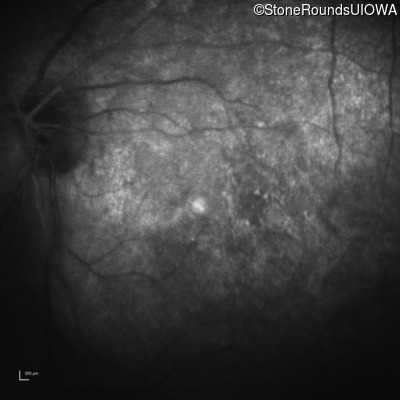
Fluorescein Angiography - Right - 20/40
Exemplar
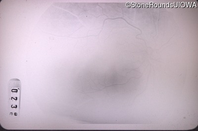
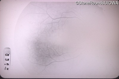
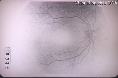
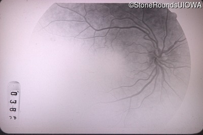
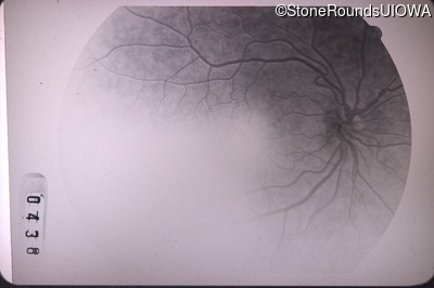
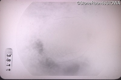
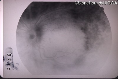
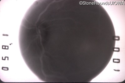
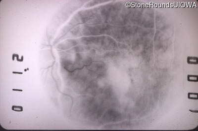
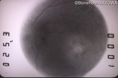
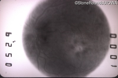
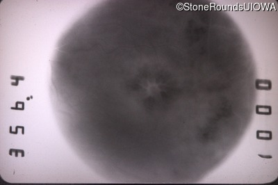
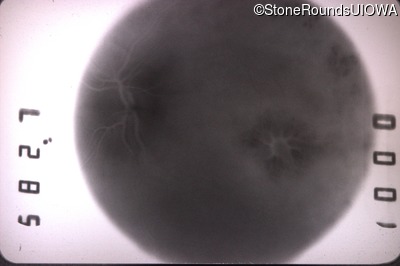
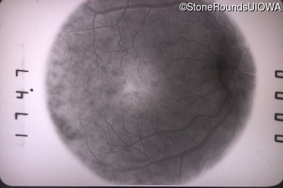
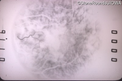
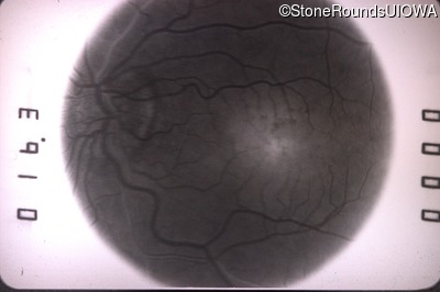
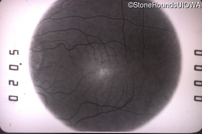
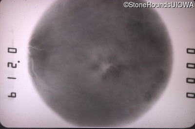
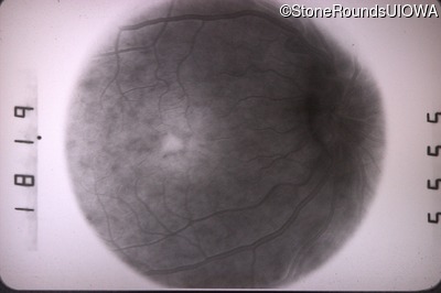
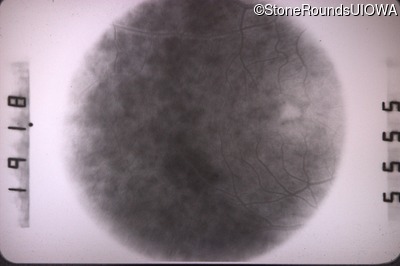
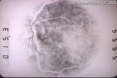
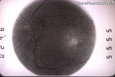
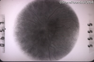
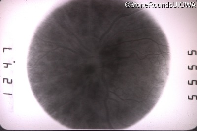
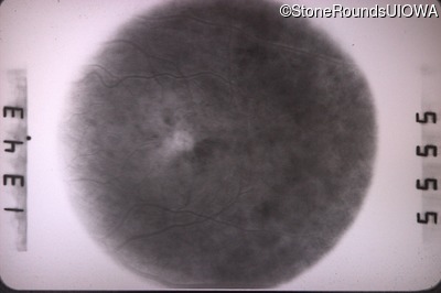
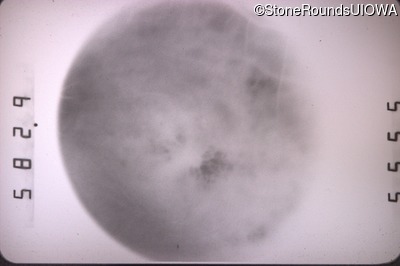
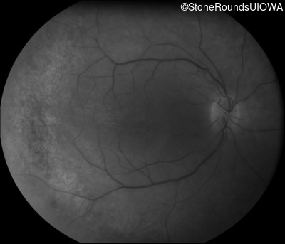
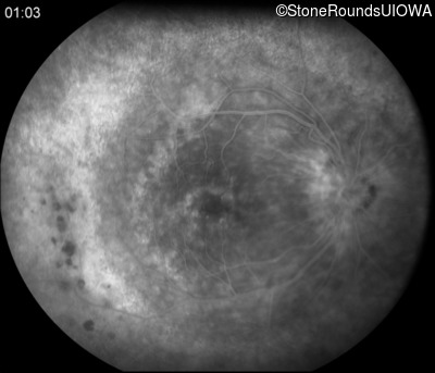
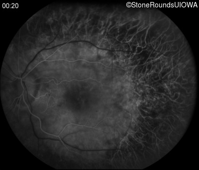
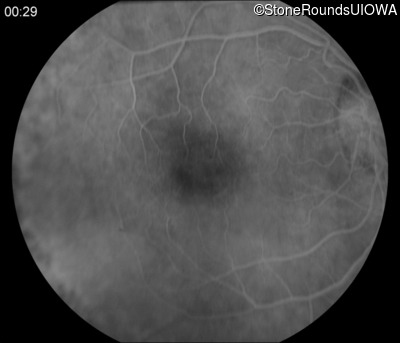
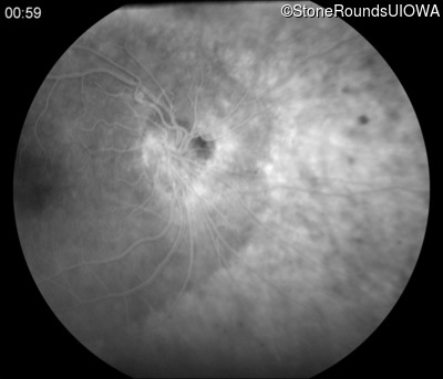
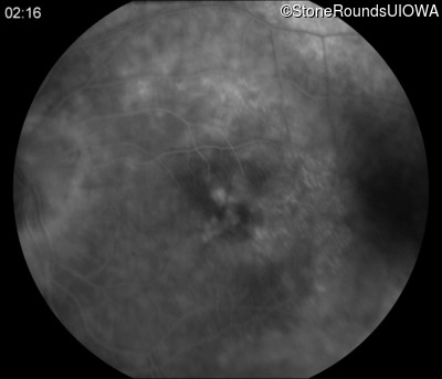
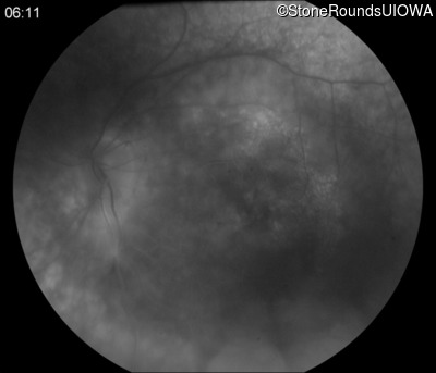
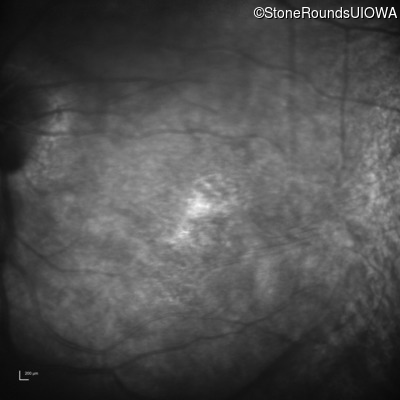
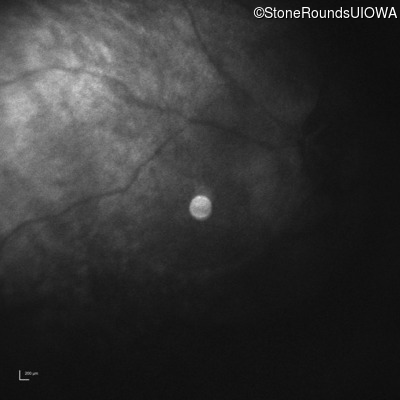
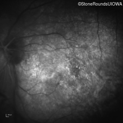
Fluorescein Angiography - Left - 20/30
Exemplar
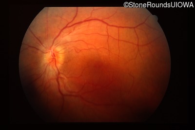
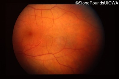
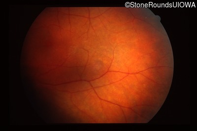
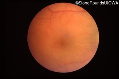
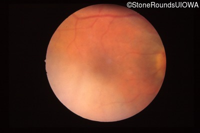
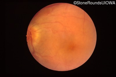
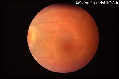
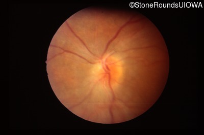
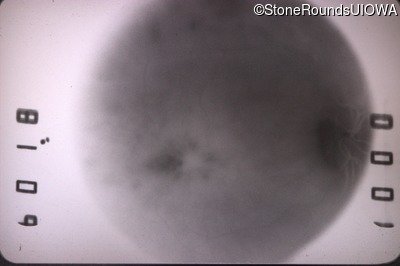
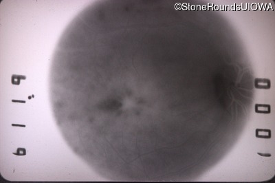
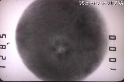
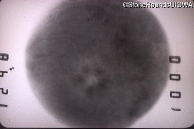
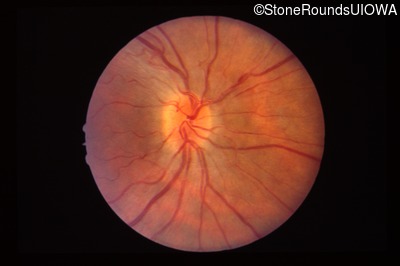
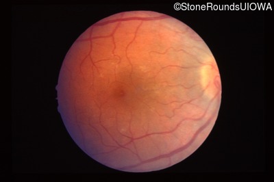
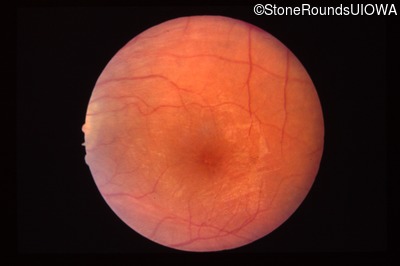
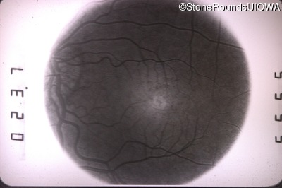
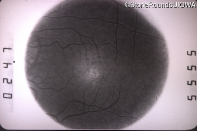
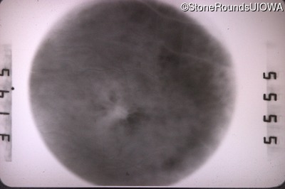
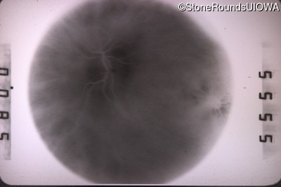
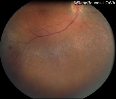
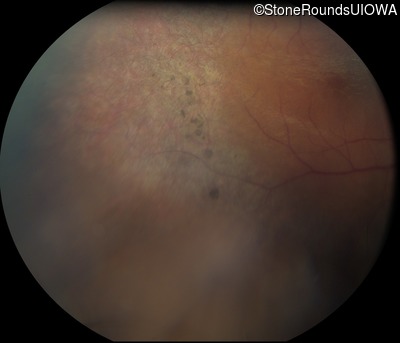
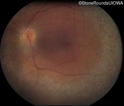
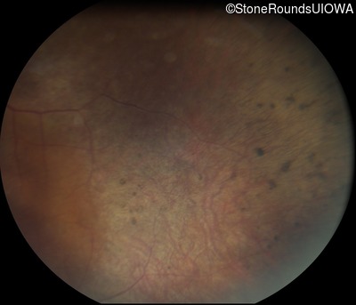
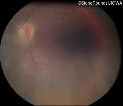
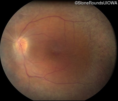
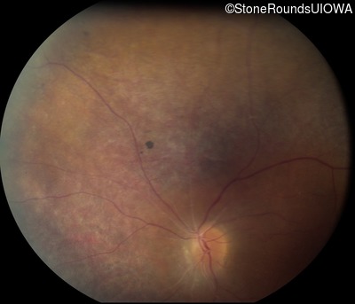
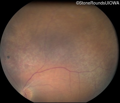
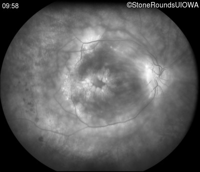
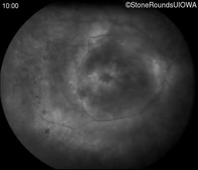
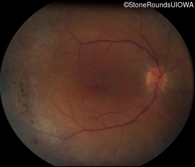
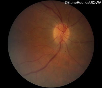
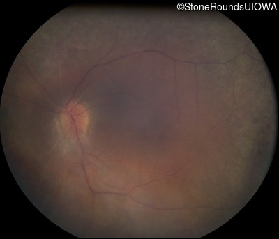
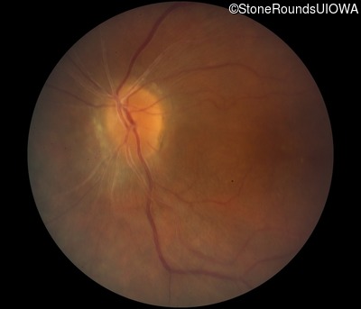
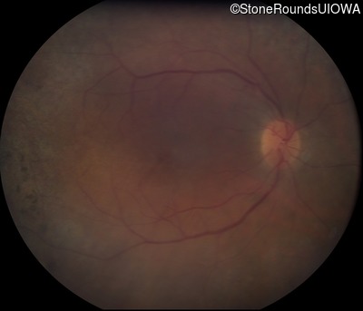
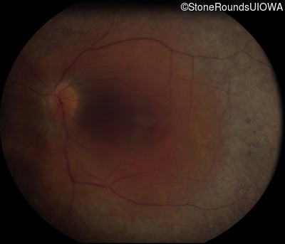
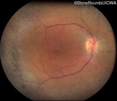
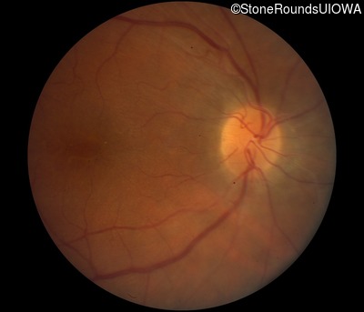
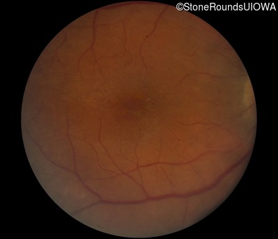
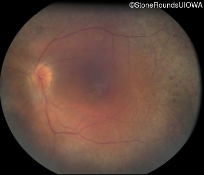
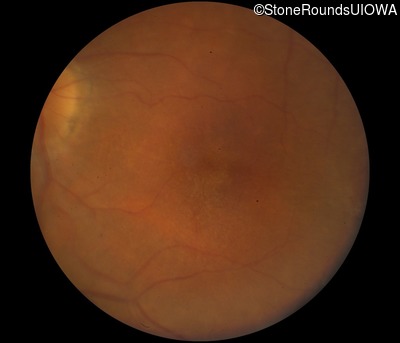
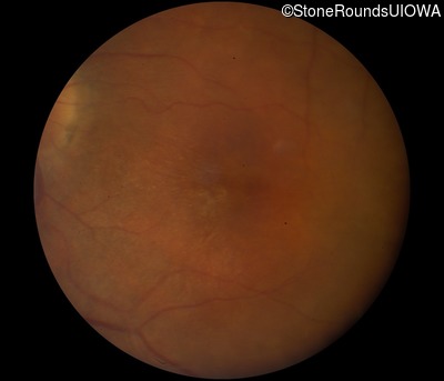
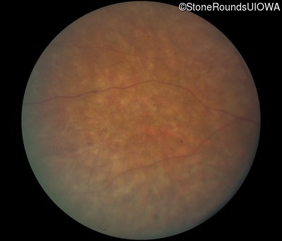
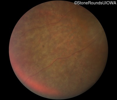
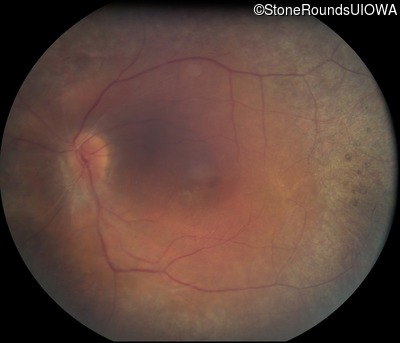
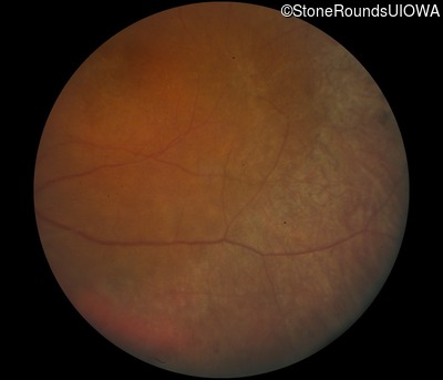
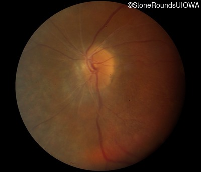
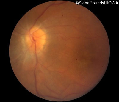
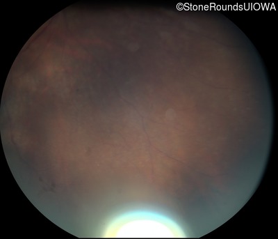
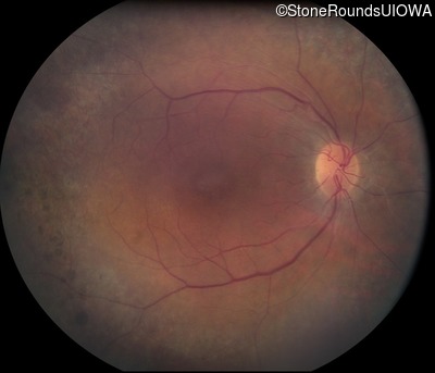
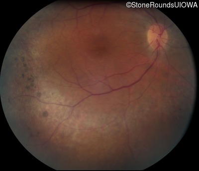
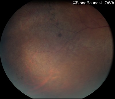
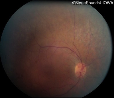
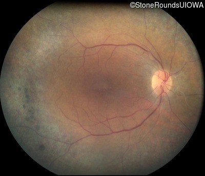
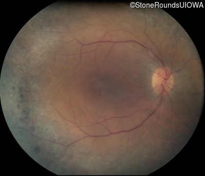
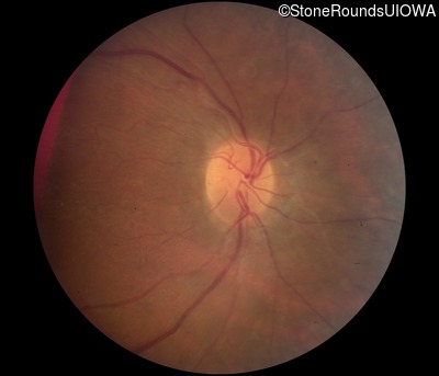
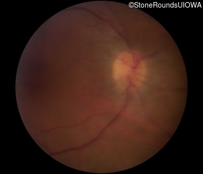
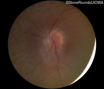
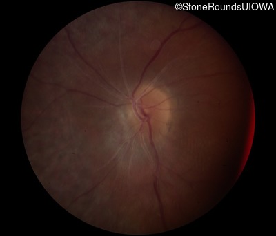
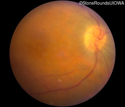
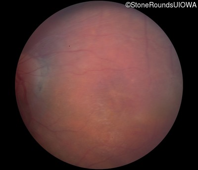
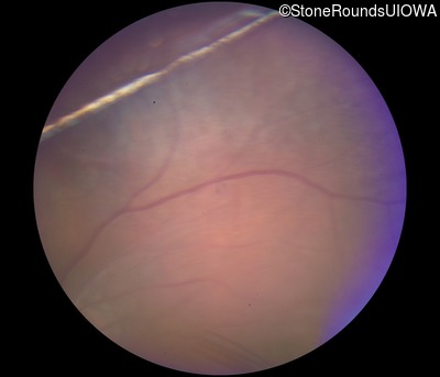
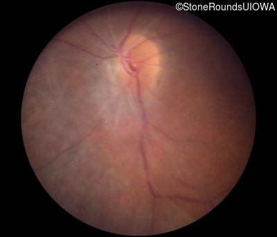
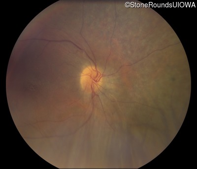
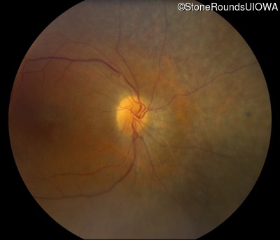
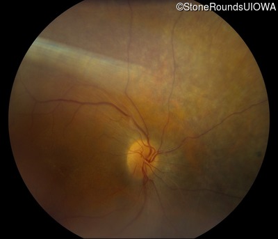
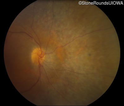
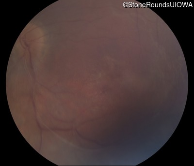
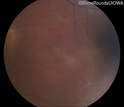
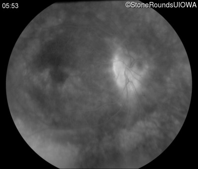
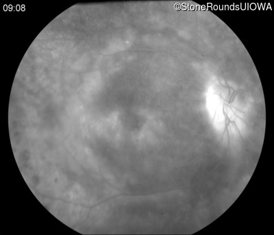
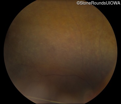
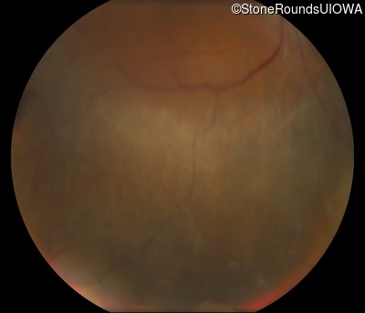
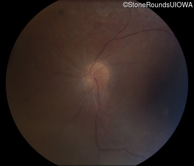
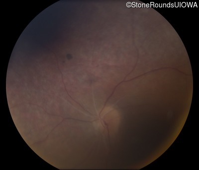
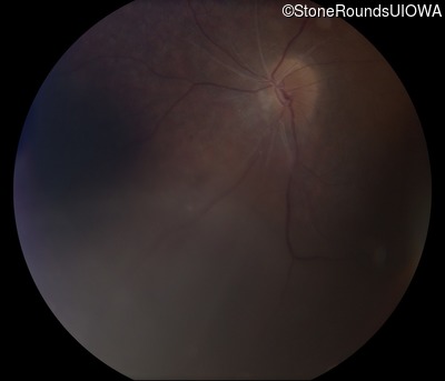
Visit at age: 25 years
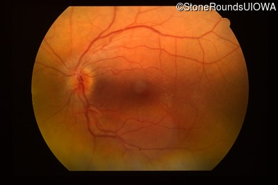
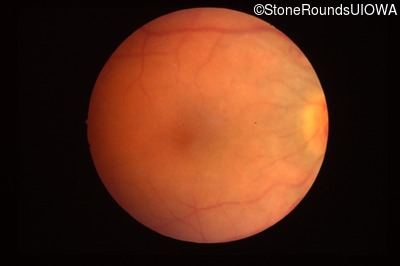
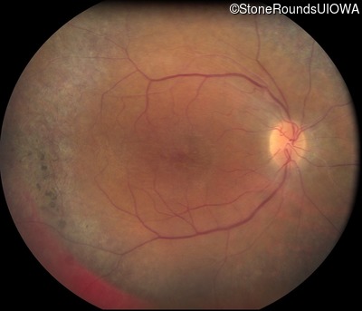
Fundus Photography - Right - 20/30
Exemplar
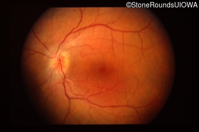
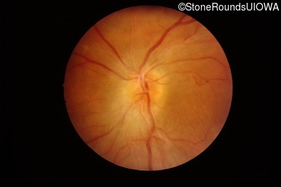
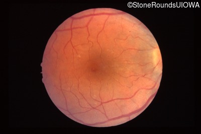
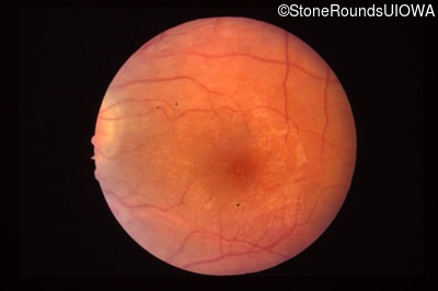
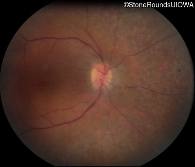
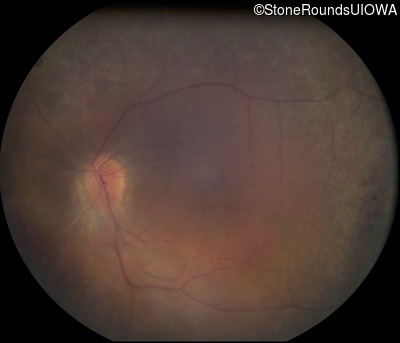
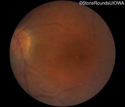
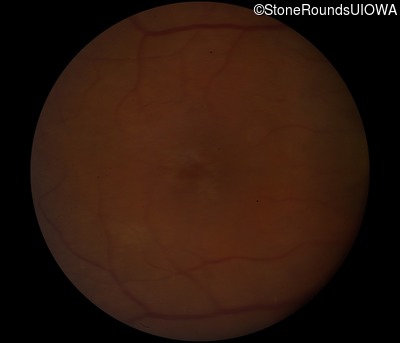
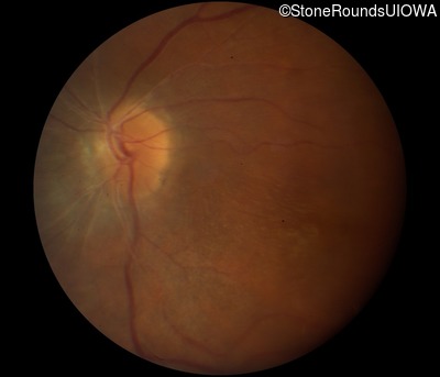
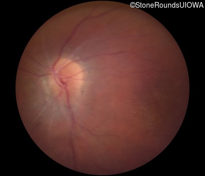
Fundus Photography - Left - 20/30
Exemplar
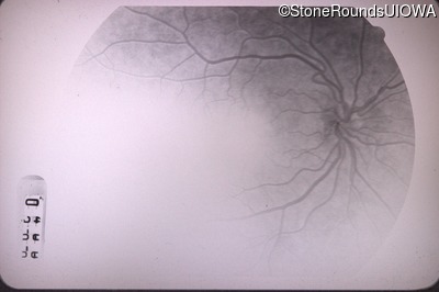
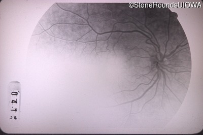
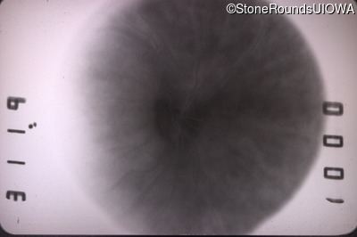
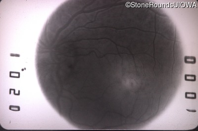
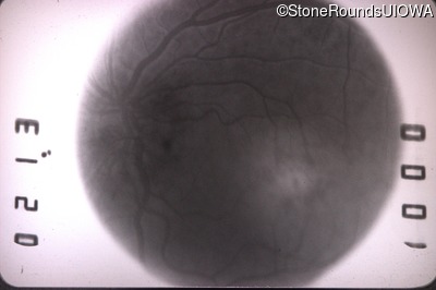
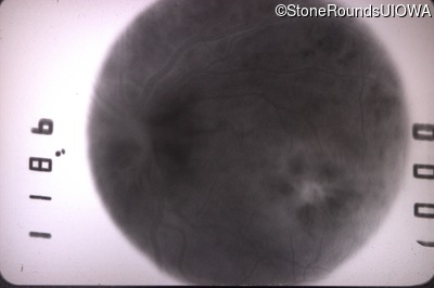
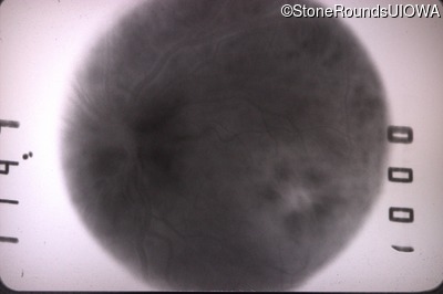
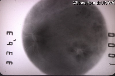
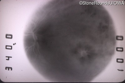
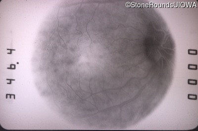
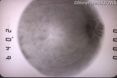
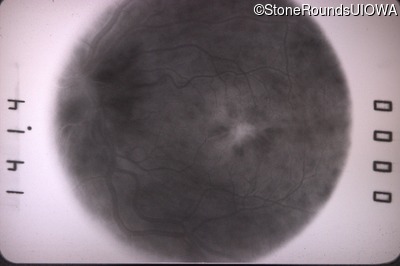
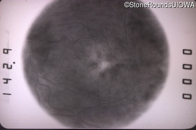
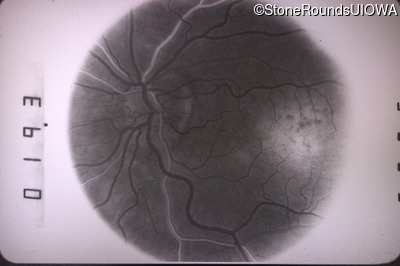
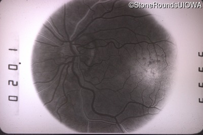
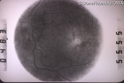
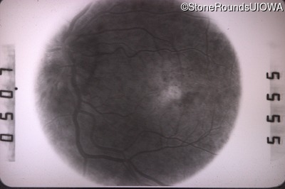
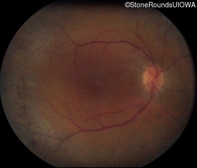
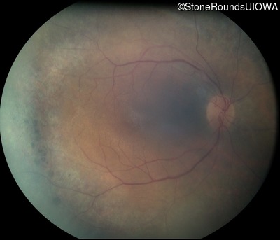
Visit at age: 28 years
Fundus Photography - Right - 20/20
Exemplar
Fundus Photography - Left - 20/50
Exemplar
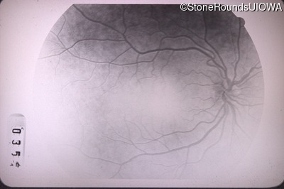
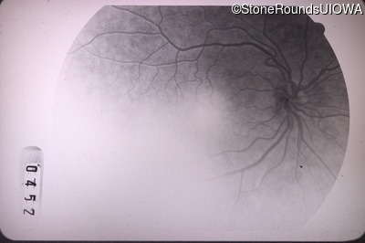
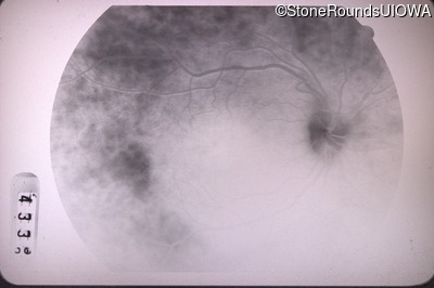
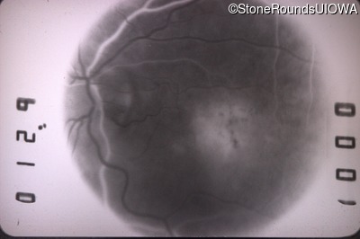
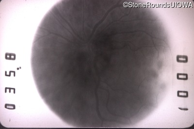
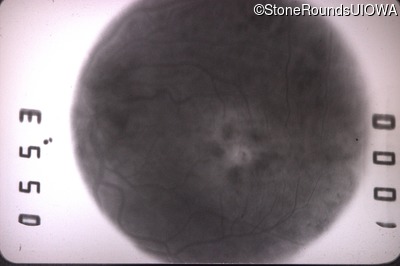
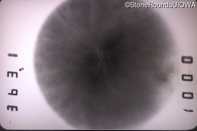
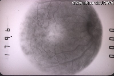
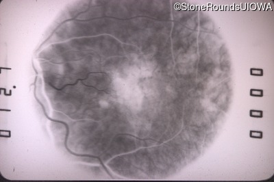
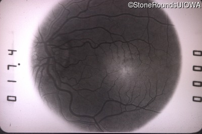
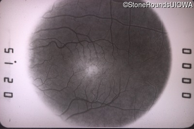
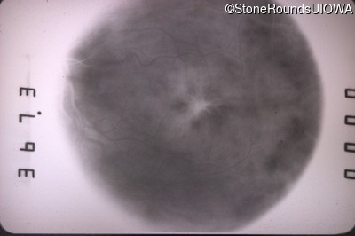
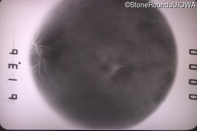
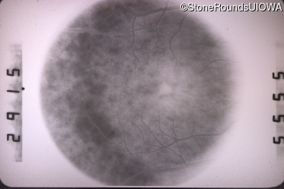
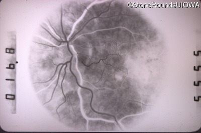
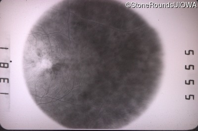
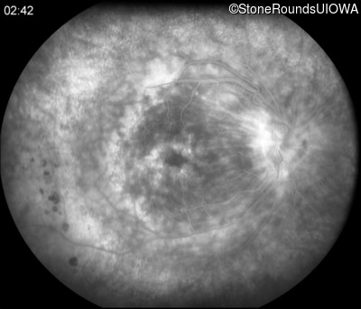
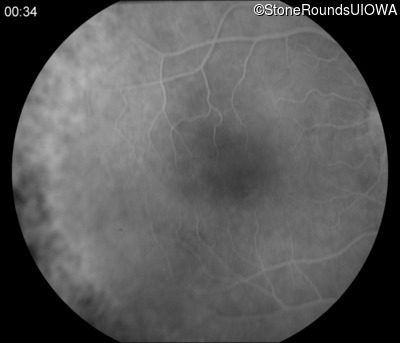
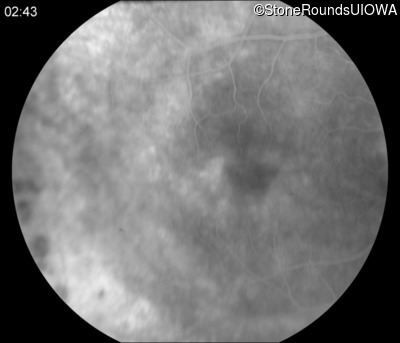
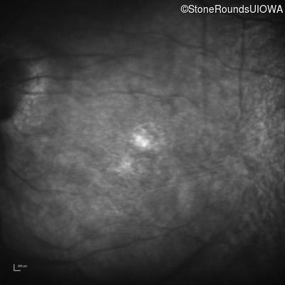
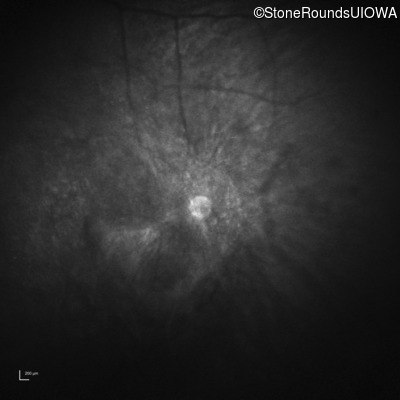
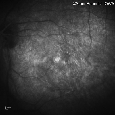
Fluorescein Angiography - Right - 20/20
Exemplar
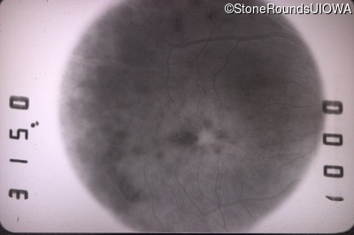
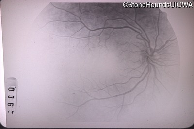
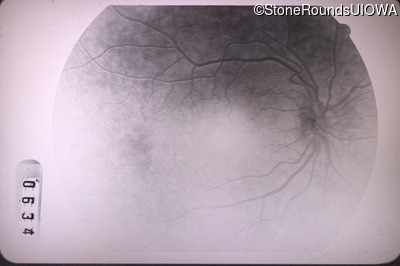
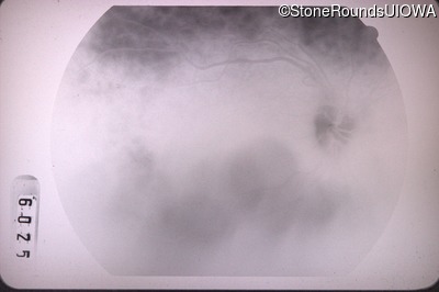
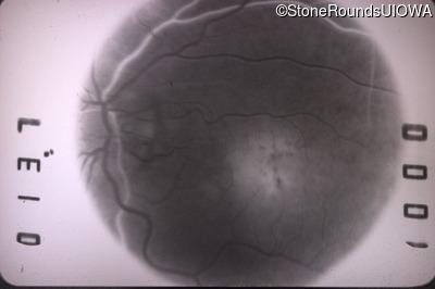
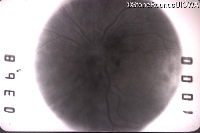
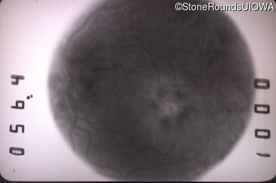
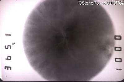
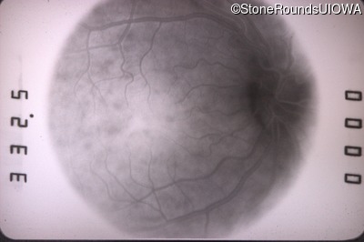
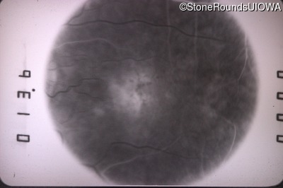
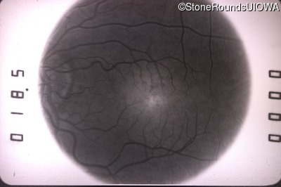
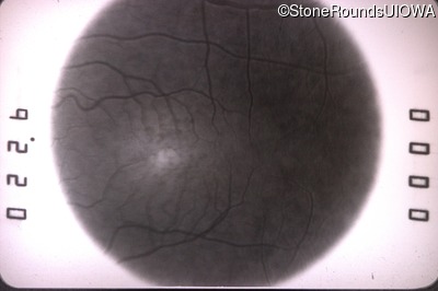
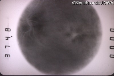
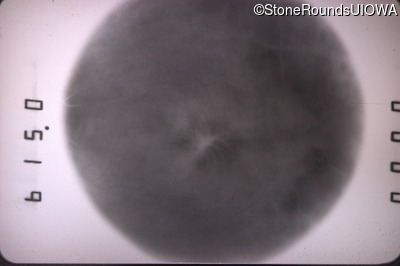
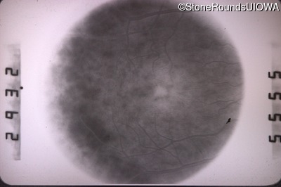
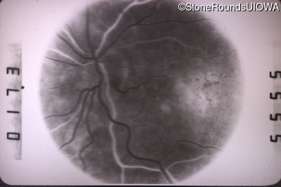
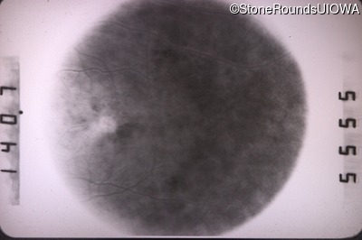
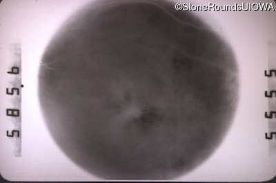
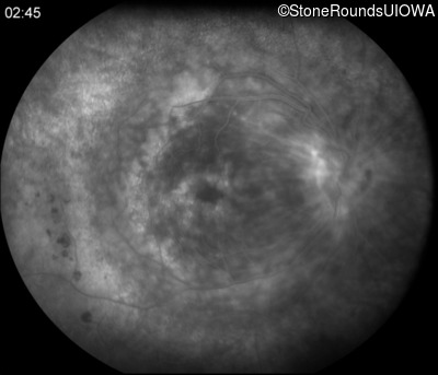
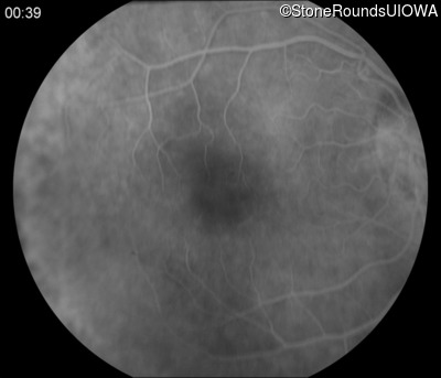
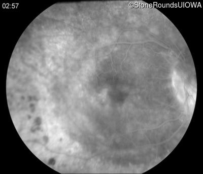
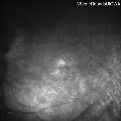
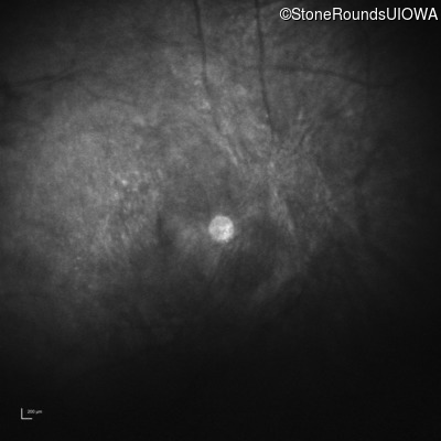
Fluorescein Angiography - Left - 20/50
Exemplar
Visit at age: 29 years
Fundus Photography - Right - 20/25 +2
Exemplar
Fundus Photography - Left - 20/40 -1
Exemplar
Fluorescein Angiography - Right - 20/25 +2
Exemplar
Fluorescein Angiography - Left - 20/40 -1
Exemplar
Visit at age: 29 years (Visit 2)
Fundus Photography - Right - 20/25
Exemplar
Fundus Photography - Left - 20/50
Exemplar
Fluorescein Angiography - Right - 20/25
Exemplar
Fluorescein Angiography - Left - 20/50
Exemplar

Visit at age: 33 years
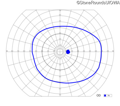
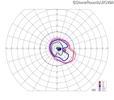
Goldmann Visual Field - Right - 20/30 +1
Exemplar
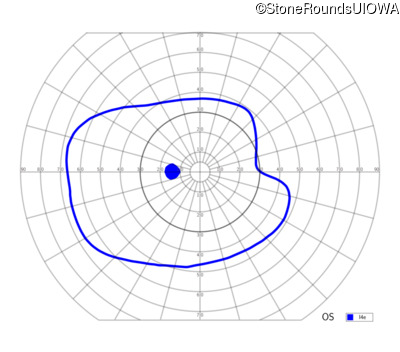
Goldmann Visual Field - Left - 20/70
Exemplar
Visit at age: 42 years
Fundus Photography - Right - 20/40 -2
Exemplar
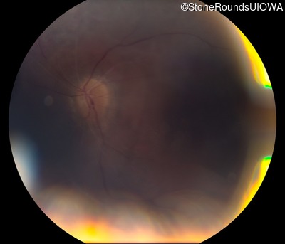
Fundus Photography - Left - 20/400
Exemplar

Fundus Montage - Right - 20/40 -2
Exemplar
Fundus Montage - Left - 20/400
Exemplar
Fluorescein Angiography - Right - 20/40 -2
Exemplar
Fluorescein Angiography - Left - 20/400
Exemplar


Visit at age: 42 years (Visit 2)
Fundus Photography - Right - 20/40 -1
Exemplar
Fundus Photography - Left - 20/300
Exemplar
Visit at age: 43 years
Fundus Photography - Right - 20/40
Exemplar
Fundus Photography - Left - 20/300
Exemplar
Visit at age: 43 years (Visit 2)
Fundus Photography - Right - 20/40
Exemplar
Fundus Photography - Left - 20/250
Exemplar
Visit at age: 43 years (Visit 3)
Fundus Photography - Right - 20/30
Exemplar
Visit at age: 44 years
Fundus Photography - Right - 20/40 -1
Exemplar
Fundus Photography - Left - 20/200
Exemplar
Visit at age: 44 years (Visit 2)
Fundus Photography - Right - 20/60
Exemplar
Fundus Photography - Left - 20/200
Exemplar
Visit at age: 45 years
Fundus Photography - Right - 20/50 +2
Exemplar
Fundus Photography - Left - 20/160
Exemplar
Fluorescein Angiography - Right - 20/50 +2
Exemplar


Fluorescein Angiography - Left - 20/160
Exemplar
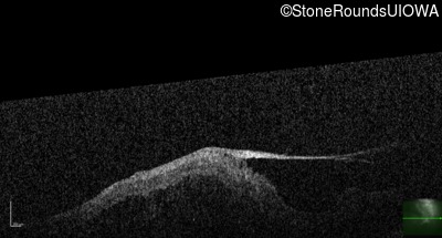
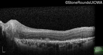
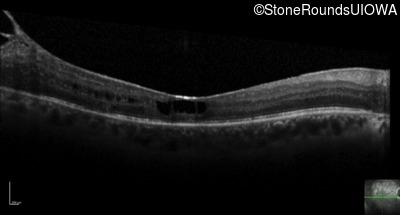
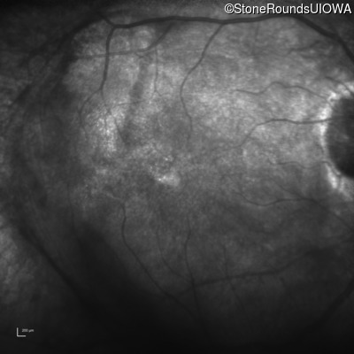
Visit at age: 45 years (Visit 2)
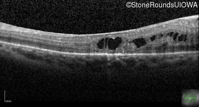
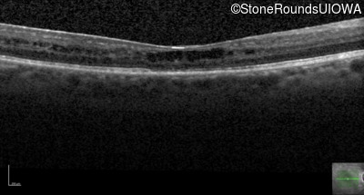
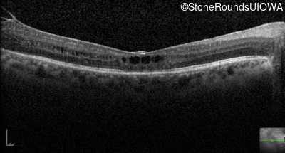
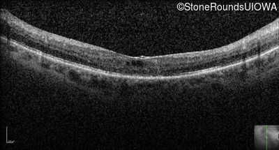
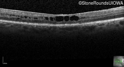
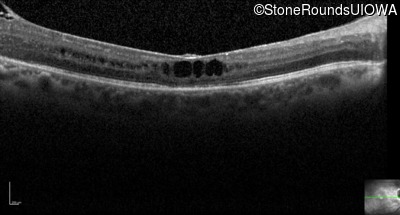
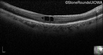
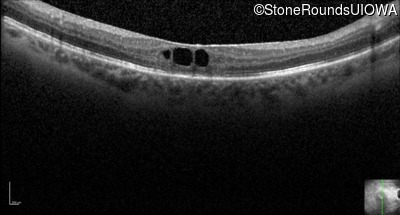
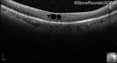
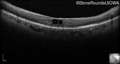
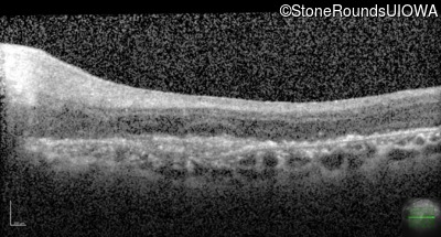
Optical Coherence Tomography - Right - 20/50 +2
Exemplar / OCT Stack
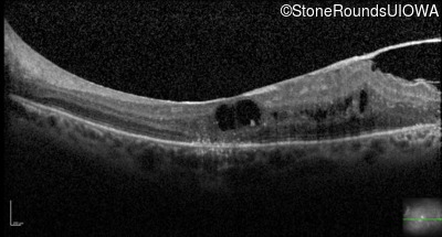
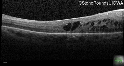
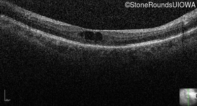
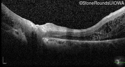
Optical Coherence Tomography - Left - 20/160
Exemplar / OCT Stack
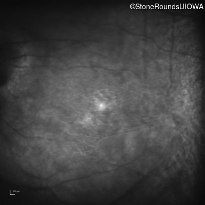
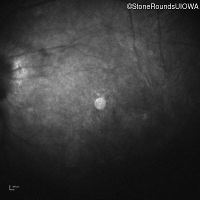
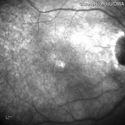
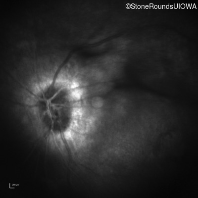
Infrared Fundus Photograph - Right - 20/50 +2
Exemplar
Infrared Fundus Photograph - Left - 20/160
Exemplar
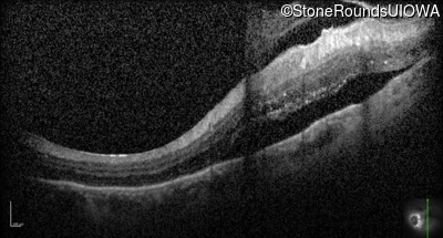
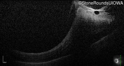
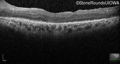
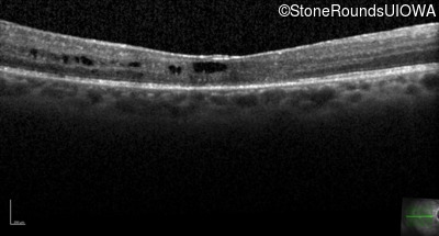
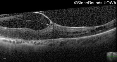
Visit at age: 46 years
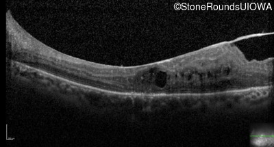
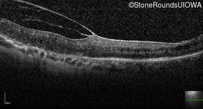
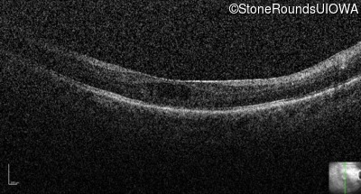
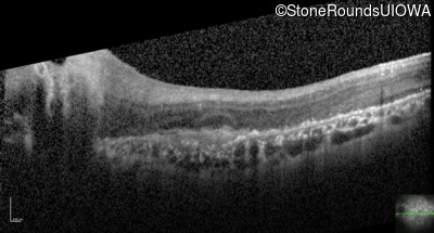
Optical Coherence Tomography - Right - 20/50 +2
Exemplar / OCT Stack
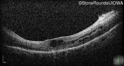
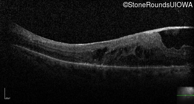
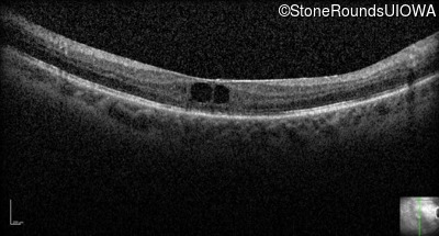
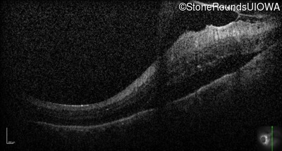
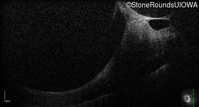
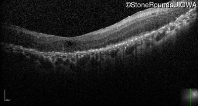
Optical Coherence Tomography - Left - 20/160
Exemplar / OCT Stack
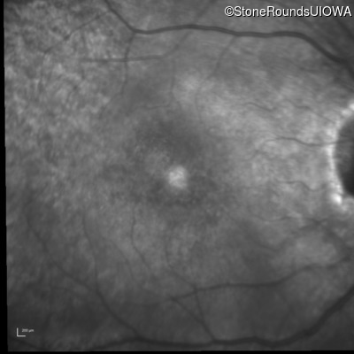
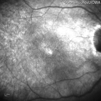
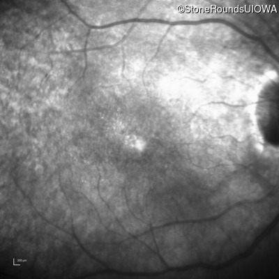
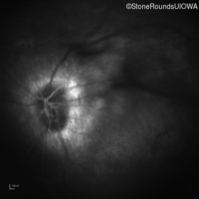
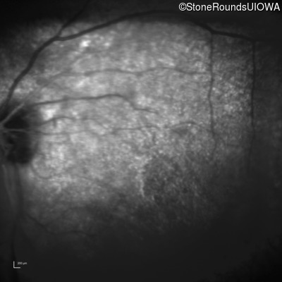
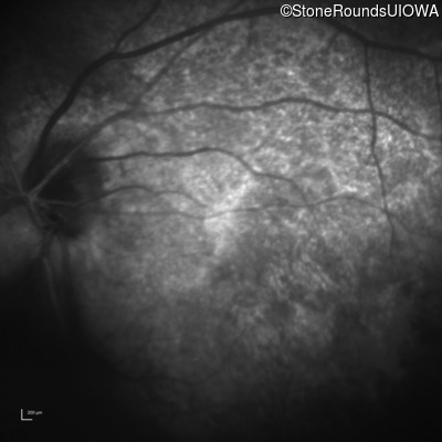
Infrared Fundus Photograph - Right - 20/50 +2
Exemplar
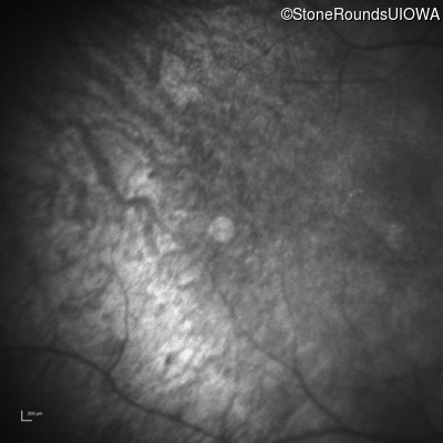
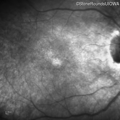
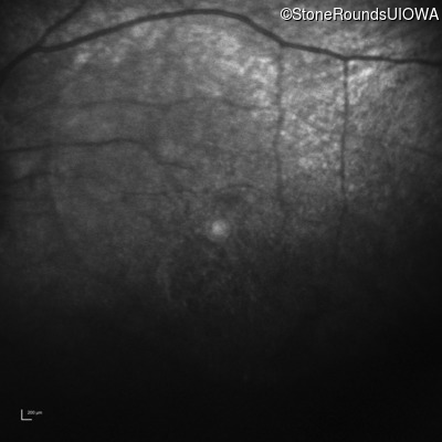
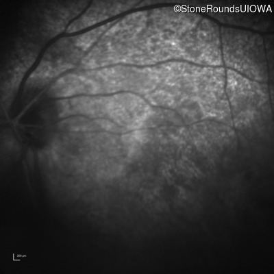
Infrared Fundus Photograph - Left - 20/160
Exemplar
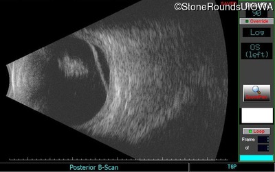
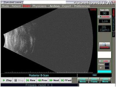
Visit at age: 46 years (Visit 2)
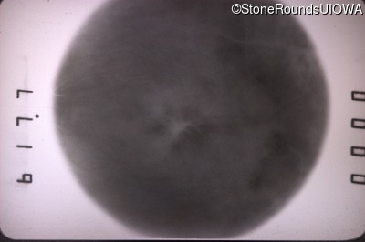
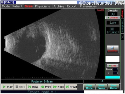
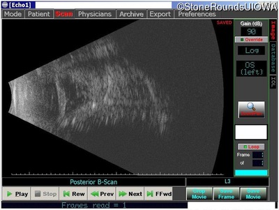
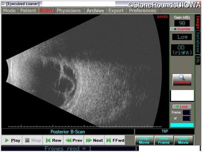
B-Scan Ultrasonography - Left - unknown
Exemplar
Visit at age: 46 years (Visit 3)
Fundus Photography - Right - 20/50 -3
Exemplar
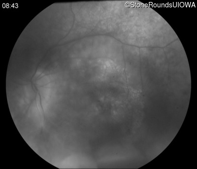
Fundus Photography - Left - Count Fingers 6"
Exemplar
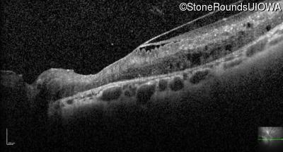
Optical Coherence Tomography - Right - 20/50 -3
Exemplar / OCT Stack

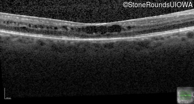
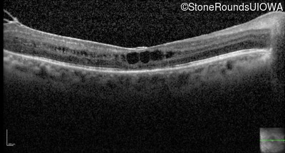
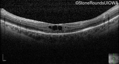
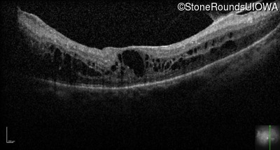
Optical Coherence Tomography - Left - Count Fingers 6"
Exemplar / OCT Stack
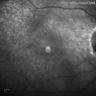
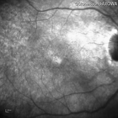
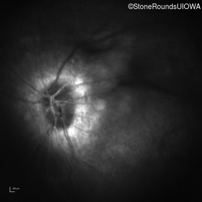
Infrared Fundus Photograph - Right - 20/50 -3
Exemplar

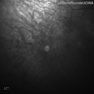
Infrared Fundus Photograph - Left - Count Fingers 6"
Exemplar
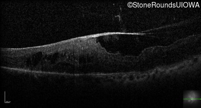
Visit at age: 46 years (Visit 4)
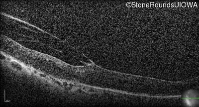
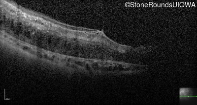
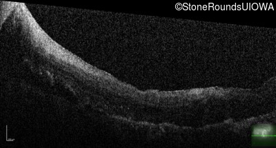
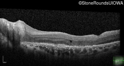
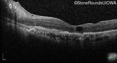
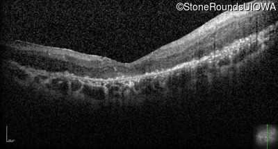
Optical Coherence Tomography - Left - Hand Motion
Exemplar / OCT Stack
OCT Stack
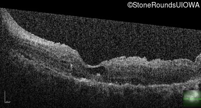
OCT Stack
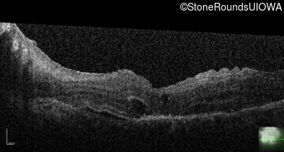
OCT Stack
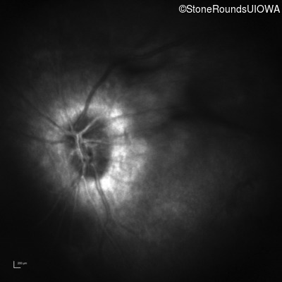
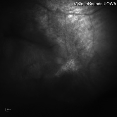
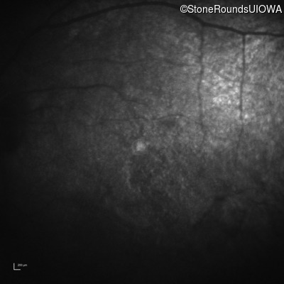
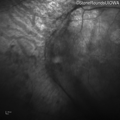
Infrared Fundus Photograph - Left - Hand Motion
Exemplar
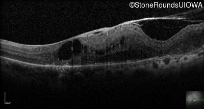
Visit at age: 46 years (Visit 5)
Fundus Photography - Left - Hand Motion
Exemplar
Optical Coherence Tomography - Left - Hand Motion
Exemplar / OCT Stack
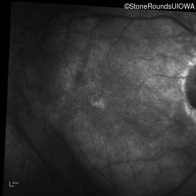
Infrared Fundus Photograph - Left - Hand Motion
Exemplar
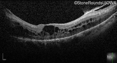
Visit at age: 46 years (Visit 6)
Optical Coherence Tomography - Left - Count Fingers 3'
Exemplar / OCT Stack
Infrared Fundus Photograph - Left - Count Fingers 3'
Exemplar
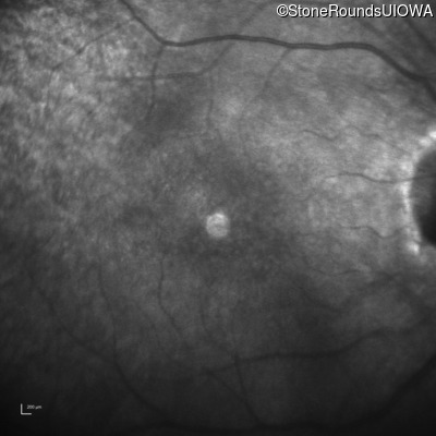
Visit at age: 46 years (Visit 7)
Optical Coherence Tomography - Left - Light Perception
Exemplar / OCT Stack
OCT Stack
Infrared Fundus Photograph - Left - Light Perception
Exemplar
Visit at age: 49 years
Goldmann Visual Field - Right - 20/70
Exemplar
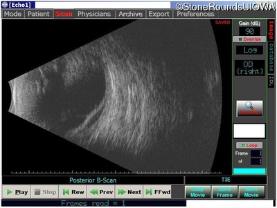
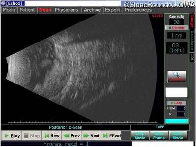
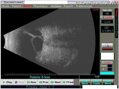
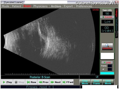
B-Scan Ultrasonography - Right - 20/70
Exemplar

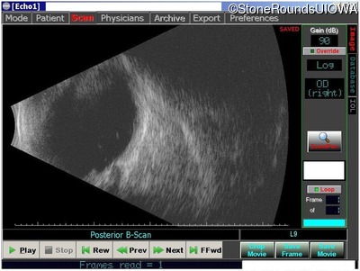
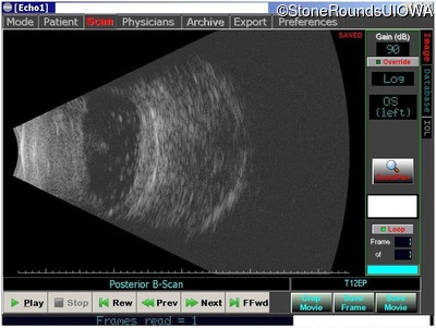
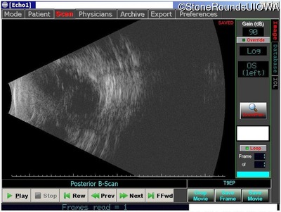
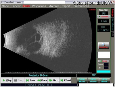
B-Scan Ultrasonography - Left - No Light Perception
Exemplar
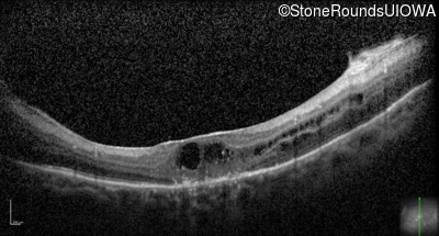
Visit at age: 50 years
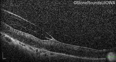
Optical Coherence Tomography - Right - 20/50 -2
Exemplar / OCT Stack
OCT Stack
OCT Stack
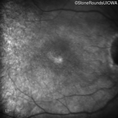
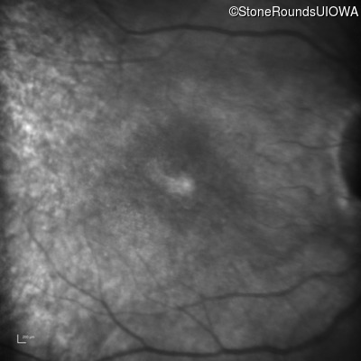
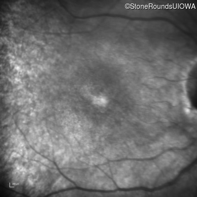
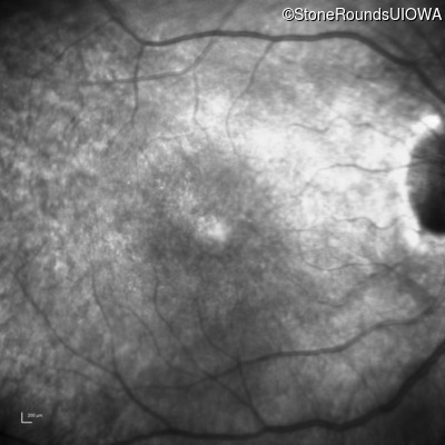
Infrared Fundus Photograph - Right - 20/50 -2
Exemplar
Visit at age: 51 years
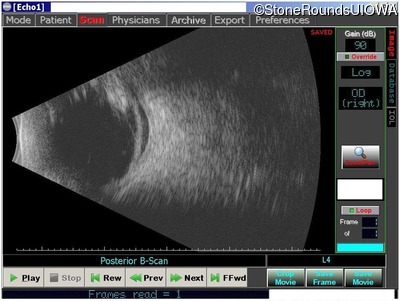
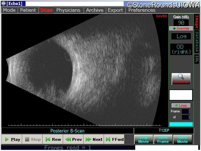
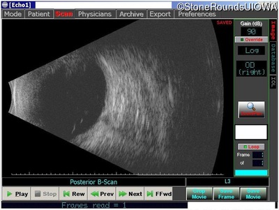
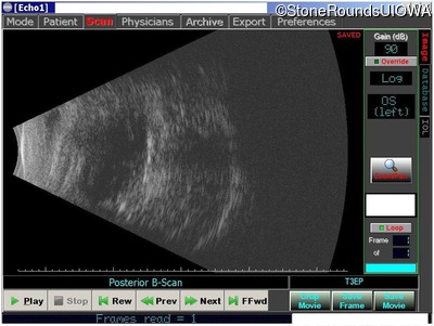
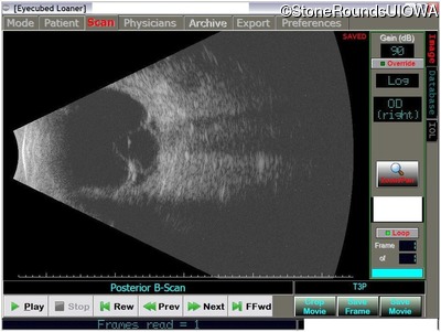
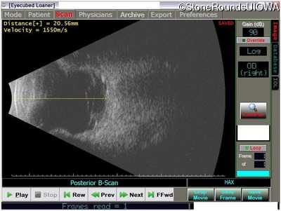
B-Scan Ultrasonography - Right - 20/400
Exemplar
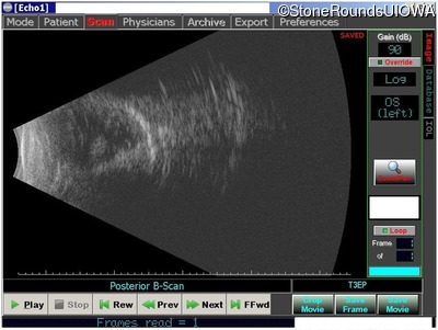
B-Scan Ultrasonography - Left - No Light Perception
Exemplar
Case Level Images